

 PDF
PDF ePub
ePub Citation
 Print
Print


The intrinsic and extrinsic carpal ligaments and triangular fibrocartilage complex (TFCC) of the wrist are major structures that guide and restrain the motion of the carpal bones. Although some injuries of the carpal ligaments may have little clinical importance, many of them can induce wrist pain or instability that often needs surgical intervention (12). Because the intrinsic carpal ligaments such as the scapholunate (SL) and lunotriquetral (LT) ligaments and the TFCC play an important role in the normal function and stability of the wrist joint, the assessment of these structures is crucial (3). Moreover, treatment options depend on the extent and location of lesions; and preoperative imaging studies, including conventional wrist arthrography, conventional magnetic resonance imaging and computed tomography or magnetic resonance arthrography (MRA), are useful not only for making the diagnosis, but also for deciding on a surgical plan (345). MRA has been widely performed instead of conventional wrist arthrography, but it is very expensive and it requires a lot of time (67).
Conventional radiocarpal arthrography was performed after radiocarpal injection under fluoroscopy guidance and several views, including wrist deviation and forearm rotation, and posteroanterior, anteroposterior, lateral and oblique views were obtained (8910). Cine arthrography takes an image every 5 seconds during intra-articular contrast injection under fluoroscopy (11).
Dynamic cine-arthrography (DCA) was performed using two processes. First, contrast injection into the radiocarpal joint through fluoroscopy guidance was done. Second, after passive wrist movement, the wrist joint was re-evaluated under fluoroscopy. This is a new technique, one which can evaluate the wrist twice under fluoroscopy.
In this study, we aimed to introduce a new technique for wrist arthrography called DCA. We compared its results with that of MRA using 1.5-T or 3-T MRI for diagnosing SL and LT ligament tears and TFCC injuries; the final diagnoses were based on arthroscopic operations.
Materials and Methods
Patient Selection
Institutional ethics review board approval was obtained and informed consent was waived. Between January, 2002 and May, 2010, a total of one-hundred forty six wrists of 141 patients with wrist pain and who underwent arthroscopic operations were initially selected for this study. Among these 146 wrists, thirty-one cases of 31 patients without DCA and seventy-seven cases of 72 patients without MRA were excluded. Finally, we enrolled in this retrospective study thirty-eight wrists of 38 patients. These wrists had undergone both DCA and MRA. All of the 38 patients were clinically suspected of having an intrinsic carpal ligament tear or a TFCC tear. There were twenty-six males and twelve females; their mean age was 36 years (range: 16-55 years). There were twenty-seven wrists on the right side and eleven on the left. The mean interval between DCA and MRA was 6 days (range: 0-67 days).
Dynamic Cine - Arthrography (DCA)
After local anesthesia was induced with about 2 mL of a 1% lidocaine solution at the dorsal surface of the wrist, we punctured the skin at the radiocarpal joint between the radius and scaphoid using a 22-gauge spinal needle under fluoroscopic guidance (SIMENSE SIREGRAPH CF, SIMENSE, Erlangen, Germany). Then, about 2-3 mL of contrast (Telebrix 30 Meglumine, Guerbet Korea, Seoul, Korea) was slowly injected into the radiocarpal joint under continuous fluoroscopy to the maximal pressure point at which no more contrast solution could be easily injected. We tried to find contrast leakage into the mid-carpal joint or the distal radioulnar joint with the wrist in a fixed pronation position during contrast injection. After intra-articular contrast injection, we did passive exercises of the wrists to the radial, ulnar, dorsal and volar directions and we re-evaluated the presence of contrast leakage via multiple direction views under fluoroscopy, including the anterior-posterior, lateral, and both oblique views. It took about 20 minutes to finish the whole process.
Wrist MR Arthrography (MRA)
Wrist MR arthrography (MRA) was done as follows: after local anesthesia with about 2 mL of a 1% lidocaine solution, a fluoroscopy-guided puncture of the radiocarpal joint between the radius and scaphoid was done using a 22-gauge spinal needle. The MR contrast solution was prepared by mixing 10 mL of normal saline and 0.2 mL of MR contrast (Megaray, Dongkook pharmaceutical, Seoul, Korea). About 1 mL of CT contrast (Telebrix 30 Meglumine, Guerbet Korea, Seoul, Korea) was injected to confirm the intra-articular puncture and then about 2-3 mL of MR contrast solution was slowly injected. MR arthrography was performed using 1.5 T or 3 T MRI. In twenty wrists, the fat-suppressed (FS) T1-weighted fast-spin echo (FSE) coronal and axial images were obtained by 1.5 T MRI (Magnetom vision plus, Siemens, Erlangen, Germany) and using a surface coil (Flex-M-coil, Siemens, Erlangen, Germany) (TR / TE, 550.0/15.0 msec for the coronal scan and 615.0/12.0 msec for the axial scan, slice thickness: 3 mm, slice gap: 0 mm, field of view: 11 cm for the coronal scan and 10 cm for the axial scan, matrix: 512 × 220, flip angle: 90°, ETL: 18 and excitations: 3). In eighteen wrists, T1-weighted spectral presaturation with inversion recovery (SPIR) coronal and axial images were taken by 3 T MRI (Achieva 3.0T X-series, Philips Healthcare, Eindhoven, The Netherlands) and using a surface coil (Sense-wrist-8, Simens, Erlangen, Germany) (TR/TE: 357.0/20.0 msec for the coronal scan and 528.0/22.0 msec for the axial scan, slice thickness: 3 mm, slice gap: 0 mm, field of view: 11 cm for the coronal scan and 10 cm for the axial scan, matrix: 316 × 313 for the coronal scan and 332 × 333 for the axial scan, flip angle: 90°, ETL: 18 and excitations: 3).
Image Analysis
The 38 cases were randomized and the radiologist was blinded to the patients' age, gender and clinical history before doing retrospective image reviews. One radiologist reviewed all of the DCA and MRA images and again he was blinded to the arthroscopic results and the findings of DCA or MRA. Evaluation of the DCA images was done first and then the MRA were investigated. The time interval of the image reviews between DCA and MRA were longer than one week.
The presence of contrast leakage into the mid-carpal joint or distal radioulnar joint was evaluated on the DCA exams. If contrast leakage into the mid-carpal joint was seen, then its location was recorded as between the scaphoid and lunate or between the lunate and triquetrum. We also recorded additional findings such as ulnar positive or negative variance, old fracture with a nonunion state of the ulnar-styloid process and subchondral sclerosis of the lunate or triquetrum indicative of chondromalacia on the scout film before intra-articular contrast injection.
On the MRA images, SL or LT ligament tears and TFCC tears were evaluated along with chondromalacia of the lunate or triquetrum. The TFCC tears were recorded based on the Palmer classification system (12, 13), that is, the TFCC tears were divided into class I traumatic injuries and class II degenerative lesions. The class I traumatic injuries were subdivided into four groups: class IA - perforation or traumatic tear of the TFC disc proper; class IB - ulnar avulsion of the TFCC with or without associated ulnar styloid fracture; class IC - distal avulsion of the TFCC through its lunate attachment (ulnolunate ligament) or its triquetrum attachment (ulnotriquetral ligament); class ID - radial avulsion at the level of the distal sigmoid notch with or without an associated sigmoid notch fracture. The class II degenerative lesions demonstrated the spectrum of ulnar impaction syndrome findings, and these were divided into five subgroups: class IIA - TFCC wear; class IIB - TFCC wear with associated lunate and/or ulnar chondromalacia; class IIC - TFCC perforation associated with lunate or ulnar chondromalacia; class IID - TFCC perforation, lunate or ulnar chondromalacia, and lunotriquetral ligament perforation; class IIE - class IID lesions with additional findings of ulnocarpal arthritis.
Arthroscopic Operation
One surgeon who was very experienced with wrist arthroscopy performed arthroscopic operations on all 38 wrists. He recorded whether there was an intrinsic ligament tear (of the SL or LT ligaments) or a TFCC tear, including chondromalacia of the lunate or triquetrum, based on the Palmer classification system. The mean interval was 57.7 days (range: 1-803 days) between DCA and the arthroscopic operation; it was 58.2 days (range: 5-803 days) between MRA and the arthroscopic operation.
Statistical Analysis
The diagnostic values (sensitivity, specificity, positive predictive value, negative predictive value and accuracy) for DCA and for MRA were calculated for SL ligament tears, LT ligament tears and TFCC tears, based on the arthroscopic findings. To evaluate the inter-examination agreement between DCA and MRA, kappa values for each tear were determined. A kappa value was considered as slight (0-0.20), fair (0.21-0.40), moderate (0.41-0.60), substantial (0.61-0.80) and almost perfect (0.81-1.00). A p value of less than 0.05 was considered to indicate a significant difference. Statistical analysis was performed using SPSS for Windows version 17.0 (SPSS Inc, Chicago, IL, USA).
Results
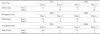
On arthroscopic operation, there were 15 SL ligament tears, 4 LT ligament tears and 28 TFCC tears among the total 38 wrists. For SL ligament tears, the sensitivity, specificity, positive predictive value, negative predictive value and accuracy were, respectively, 66.7% (10/15), 100% (23/23), 100% (10/10), 82.0% (23/28) and 86.8% (33/38) for DCA; for MRA they were, respectively, 80% (12/15), 95.7% (22/23), 92.3% (12/13), 88% (22/25) and 89.5% (34/38) for MRA. For LT ligament tear, the sensitivity, specificity, positive predictive value, negative predictive value and accuracy were 75.0% (3/4), 94.1% (32/34), 60.0% (3/5), 97.0% (32/33) and 92.1% (35/38) for DCA, and they were 75.0% (3/4), 91.2% (31/34), 50.0% (3/6), 96.9% (31/32) and 89.5% (34/38) for MRA, respectively. For TFCC tears, the sensitivity, specificity, positive predictive value, negative predictive value and accuracy were 96.4% (27/28), 100% (10/10), 100% (27/27), 90.9% (10/11) and 97.4% (37/38) for both DCA and MRA, respectively (Table 1). There was no significant difference between the results for TFCC tears. (Chi-square test, p > 0.05).
For the diagnosis of SL ligament tears, DCA and MRA demonstrated almost perfect inter-examination agreement (kappa value = 0.814). There was also perfect inter-examination agreement between DCA and MRA for TFCC tears (kappa value = 1.000). For LT ligament tears, the inter-examination agreement between DCA and MRA was substantial (kappa value = 0.682) (Table 2).
Regarding the Palmer classification, there were ten class IA lesions, one class IB lesion and three class ID lesions of a total of 28 TFCC tears on arthroscopic operations. For the rest, twelve class IIC lesions (with lunate/ulnar chondromalacia) and two class IID lesions (with lunate/ulnar chondromalaia and LT ligament tear) were found. In addition, one class IIA lesion (TFCC wear) and six class IIB lesions (TFCC wear with lunate/ulnar chondromalacia) were seen on arthroscopic operations, and were not included as TFCC tears because there was no perforation.
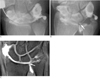
The Palmer classification results were equal for both DCAs and MRAs in half of the 28 TFCC tears (14/28, 50%). All of them were confirmed on arthroscopic operation: nine class IA lesions, three class ID lesions, one class IIB lesion and two class IIC lesions. Only one class IA lesion was diagnosed correctly as a class IA lesion on DCA, but it was thought to be a class IID lesion on MRA, indicating a false-positive LT ligament tear (1/10, 10%). Nine class IIC lesions were mistaken for class IA lesions on DCA (9/12, 75.0%), of which five were misinterpreted as class IA lesions on MRA (Fig. 1). One class IIC lesion was regarded as a class IID lesion on MRA; it showed a false-positive lunate chondromalacia. A total of six class IIC lesions were not diagnosed correctly on MRA (6/12, 50%). Both the diagnoses of the class IID lesions diagnosed on arthroscopy were considered faulty as one was a class IA lesion on DCA (Fig. 2) and the other was a class IIC lesion with marked ulnar positive variance on DCA (2/2, 100%); one was seen as a class IIC lesion on MRA (1/2, 50%). As for the one class IB lesion and the one IIA lesion diagnosed on arthroscopy, neither DCA nor MRA was accurate as neither modality noted any abnormality around the TFCC (1/1, 100% for class IB and IIA lesions). Meanwhile, five out of six class IIB lesions were regarded as having no abnormality of the TFCC on DCA (5/6, 83.3%) and two of them were mistaken for class IIA lesions (2/6, 33.3%).
Discussion
A TFCC tear is a common cause of pain and instability of the wrist. A TFCC tear may not be apparent on a clinical examination alone, and a variety of radiographic tools have been used to enhance the diagnostic accuracy. Imaging studies of the carpal ligaments have also played an important role in the assessment of patients complaining of wrist pain (514).
Conventional direct arthrography is a well-established diagnostic technique for the evaluation of wrist pain, and the advanced forms of arthrography such as cinefluorography have allowed the techniques of diagnostic arthroscopy to be implemented at the wrist joint (1315).
Diagnosing a TFCC tear by direct MRA has not been
entirely satisfactory, although the relatively high positive predictive value for MRA has been reported for the detection of TFCC tears. The negative results of MRA in patients with a clinical suspicion of an TFCC tear should be interpreted with caution (16). On the other hand, conventional direct arthrography has been regarded as being effective for evaluating TFCC tears (17). However, if contrast is seen at the mid-carpal joint, it is often difficult to determine which intercarpal ligament is torn: the SL or the LT ligament. Therefore, we assumed that DCA could be helpful for evaluating the intercarpal ligament or TFCC by continuous fluoroscopic evaluation, especially for the evaluation of LT and SL ligament injuries. Because contrast leakage into the mid-carpal joint may develop during or after passive wrist movement on DCA, partially-healed intercarpal ligament tears or flap tears are expected to be more detectable because they show delayed contrast leakage into the mid-carpal joint or distal radioulnar joint. DCA not only may provide better delineation of the carpal ligaments, but also, it demonstrates the presence or absence of contrast leakage through the carpal ligament tears (11).
A single-injection of the radiocarpal joint alone may be sufficient during wrist arthrography instead of the traditional triple-injection wrist arthrography (1819). Single-injection wrist arthrography is superior to routine MRI for the detection of full-thickness TFCC tears (20). In this study, we used a single-compartment injection via the radiocarpal joint for both DCA and MRA, and this demonstrated pretty good diagnostic performances, especially for TFCC tears.
In this study, the diagnostic accuracy of DCA was similar with that of MRA for SL ligament and LT ligament tears (86.8% of DCA and 89.5% of MRA for SL ligament tear; 92.1% of DCA and 89.5% of MRA for LT ligament tear). For TFCC tears, the overall accuracy of both was same (97.4% for both DCA and MRA). The inter-examination agreements between DCA and MRA had relatively high kappa values (from substantial to perfect), and this is indicative of good inter-examination agreement.
Regarding the Palmer classification, there were many mismatching cases between DCA and MRA. Nine cases of Palmer class IIC lesions (9/12, 75%) were mistaken for class IA lesions on DCA because chondromalacia of the lunate or triquetrum was difficult to find on DCA, except for the cases with severe joint space narrowing. Six Palmer class IIC lesions (6/12, 50%) could not be exactly diagnosed on MRA because of the limitation for evaluating chondromalacia or LT ligament tears. Therefore, chondromalacia of the lunate or triquetrum may be hard to determine correctly on both DCA and MRA. For the diagnosis of Palmer class IIB lesions, MRA may be superior to DCA, as the latter resulted in five false-negative cases. Because the Palmer classification result is not a main factor that influences the treatment planning, this misinterpretation of DCA may not be important in actual clinical practice.
This study has some limitations. First, only full-thickness tears of the TFCC, SL and LT ligaments on arthroscopy were considered positive lesions. Less well defined lesions (e.g., partial-thickness tears in the TFCC and intercarpal ligaments) were not included in the determination of the sensitivity, specificity or accuracy of MRA. Second, the radiation dose by continuous fluoroscopy was not measured. So, we can not recommend an optimal examination time of DCA for detecting delayed contrast leakage.
In conclusion, DCA with a single-compartment injection may be more helpful for the evaluation of wrist pain or instability instead of MRA, especially for TFCC tears.

 XML Download