

 PDF
PDF ePub
ePub Citation
 Print
Print


Primary cutaneous lymphomas are defined as a subtype that are restricted to the dermal or epidermal region and are without any signs of systemic lymphoma. Most skin non-Hodgkin's lymphomas (NHL) originate from T cells, and primary cutaneous diffuse large B cell lymphomas (PCDLBCLs) are uncommon (1). PCDLBCLs appear clinically as non-specific multiple palpable nodules or plaques in the subcutaneous tissue (2). PCDLBCLs localized in the head and trunk have good outcome compared to PCDLBCLs involving the leg (34). The clinical and pathological features of PCDLBCLs have been well described, but MR imaging features of PCDLBCLs have not been reported. Herein we report such features.
Case Report
A 67-year-old woman presented with fever and a subcutaneous mass on the left posterior chest wall. She had had a small non-tender palpable nodule on her left posterior chest wall for 20 years, and it had recently increased in size. Palpable lymphadenopathy or organomegaly was not found on physical examination. Laboratory tests disclosed anemia (a hemoglobin level of 10.8 g/dL), but white blood cell and platelet counts were within the normal ranges.
To diagnose the mass and assess its extent, MRI was performed using a 1.5 T MR system (Symphony Vision®, Siemens Medical Solutions, Erlangen, Germany). MR images included T2-weighted turbo spin echo sequences (TR/TE, 3700-4400/99 msec; echo train length, 13; 4 mm slice thickness; 448 × 252 matrix; 20 cm field of view) and T1-weighted turbo spin echo (TR/TE, 700/11 msec; 448 × 233 matrix; 4 mm slice thickness; 20 cm field of view) sequences. Three minutes after injection of contrast material (0.1 mmol/kg at a rate of 2-3 mL/sec; Gd-diethylenetriaminepentaacetic acid, Magnevist; Bayer Schering Pharma, Berlin, Germany), post-contrast images were obtained using spin echo sequences (TR/TE=640/11 msec) with a fat suppression technique.
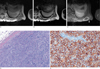
T1-weighted images showed a 3 × 2.5 × 2 cm, lobulated subcutaneous mass of low signal intensity with a high signal rim and infiltration of the overlying skin (Fig. 1A) in her left posterior chest wall. T2-weighted images showed a hyperintense mass with thin septa (Fig. 1B). On gadolinium-enhanced, fat-suppressed, T1-weighted images, a thick irregular peripheral rim and septal enhancement were seen. Fuzzy enhancement of overlying skin was also seen (Fig. 1C). The differential diagnoses included a ruptured epidermoid cyst, a cutaneous abscess, and, less likely, solid mass.
On general anesthesia, excisional biopsy was performed. A yellowish, 2 cm diameter, soft, and movable mass was seen in the subcutaneous layer of the left posterior chest wall. It was easily separated from adjacent soft tissue.
A section of the excisional biopsy revealed a yellowish mass with central atrophy and a peripheral myxoid component. Microscopic findings of the mass disclosed a dense and diffuse infiltration of atypical lymphocytes in the dermis and subcutaneous tissue (Fig. 1D). An immunohistochemical study showed positivity of lymphocytes for CD20, CD99, CD10, BCL6 and Ki-67 LI (Fig. 1E). However, tumor cells were negative for CD3, CD30, BCL2, EMA, CD56, Cytokeratin (AE1/AE3), Cyclin D1, ALK and MUM 1. Combined morphologic and immunohistochemical features supported a diagnosis of diffuse large B-cell lymphoma. To assess extracutaneous manifestations of lymphoma, CT of the chest, abdomen, and whole-body F-18-deoxyglucose positron emission tomography (FDG-PET) were performed. Neither abnormal lymphadenopathy nor visceral involvement was noted. The patient has been periodically monitored in our hospital over the last 6 months without any recurrence of the lymphoma.
DISCUSSION
Nearly half of NHLs arise in extranodal sites, most commonly affecting the gastrointestinal tract and skin. The annual incidence of primary cutaneous lymphomas (PCLs) is 0.5 to 1 per 10,000 people (5, 6). Mycosis fungoides is the most common subtype of PCLs in the West. It is thought that 20 to 25% of these cases are of B-cell origin. PCDLBCLs represent a heterogeneous group of NHLs that present in the skin without evidence of extracutaneous disease at presentation and within 6 months after diagnosis. In the most recent (2005) classification of the World Health Organization/European Organization for Research and Treatment of Cancer, four main types of PCDLBCLs are recognized: primary cutaneous marginal zone B-cell lymphoma, primary cutaneous follicle-center lymphoma, primary cutaneous diffuse large B-cell lymphoma-leg type and primary cutaneous diffuse large B-cell lymphoma, other (7). PCDLBCLs usually affect elderly patients, in particular females, and characteristically present as solitary or multiple persistent red to violaceous papules, plaques, or non-scaly, non-ulcerated smooth cutaneous nodules on the head, trunk and leg. Therefore, PCDLBCLs are often confused with other entities including subcutaneous inflammatory panniculitis, cystic masses, solid tumors and vascular lesions (68).
Imaging tools such as ultrasound (US), computed tomography (CT), FDG-PET and MRI have an advantage over physical examination for detection and follow up (34). US is useful for evaluating a superficially located lesion. However, it is generally used for regional evaluation rather than to evaluate the extent of disease. CT has been useful in staging the lymphoma and in defining the extent of adenopathy. Although it is associated with limited soft tissue delineation and radiation, CT is still the first choice for soft tissue evaluation. FDG-PET can detect most low-grade lymphomas, but some types of indolent lymphoma are not well visualized on FDG-PET. Moreover, the spatial resolution of FDG-PET is not perfect: lesions smaller than 1 cm in diameter often go undetected. However, further studies are needed to assess the diagnostic ability of FDG-PET to detect PCDLBCLs. MRI is the radiologic technique of choice for preoperative evaluation of the extent and depth of primary and recurrent skin tumors.
The key point that enables the diagnosis of primary extranodal soft tissue lymphoma to be made is the extension of the tumor with preservation of the surrounding structures. In this patient, muscles and ribs were closely contacted by, but not invaded by the lymphoma, which is not a specific finding. On MR imaging, extranodal soft tissue lymphomas are homogeneously isointense or slightly hypointense relative to normal muscle on T1-weighted spine echo images, and are hyperintense to muscle on T2-weighted spin echo images. They uniformly enhance after intravenous administration of contrast material (9). Our case showed a well-defined lobulating lesion of high signal intensity on T2-weighted images, and low signal intensity with a peripheral high signal intensity rim on T1-weighted images in the subcutaneous layer of the chest wall. After gadolinium enhancement, we observed a central non-enhancement area with a thick irregular peripheral rim enhancing nodule and patchy enhancement of overlying skin. In correlation with the histopathology, the peripheral rim reflected a myxoid tissue, and the central area reflected necrosis and atypical lymphocytic infiltration of overlying skin. MR imaging features of PCDLBCLs are not definitively diagnostic because benign inflammatory lesions such as abscesses, complicated ganglia, bursitis, and ruptured epidermoid cysts also show septa and demonstrate thick and irregular rim enhancement after gadolinium enhancement (10).
A cutaneous T-cell lymphoma generally appears as eczema-like skin rashes or plaque-like lesions affecting any part of the body. Unlike cutaneous T cell lymphoma, cutaneous B cell lymphoma often shows nodule-like lesions (2). Imaging features of cutaneous lymphoma are non-specific and include soft tissue thickening, infiltration, or a mass. MR imaging features of PCDLBCLs have not been reported.
Inclusion of extranodal soft tissue lymphoma in the clinical differential diagnosis affects surgical decision making because lymphoma is not treated with surgery. In general, complete resection is not indicated because it would require the removal of a marker of disease response to chemotherapy, radiation therapy, or both.
In conclusion, PCDLBCL is a rare disease that can have various imaging features. Although a soft tissue mass with central necrosis and a thick enhanced rim in the subcutaneous layer and infiltrating overlying skin may simulate a benign inflammatory lesion or a benign neoplasm on MR imaging, awareness of PCDLBCLs by clinicians and radiologist may help to achieve an accurate differential diagnosis.
 XML Download