

 PDF
PDF ePub
ePub Citation
 Print
Print


Fenestrations or duplications of the internal jugular vein (IJV) are a rare congenital anomaly in the neck. Although many arterial fenestrations have been reported in the vertebral artery, basilar artery, middle cerebral artery, anterior cerebral artery, posterior inferior cerebellar artery, posterior cerebral artery, posterior communicating artery, and internal carotid artery, fenestration of the IJV is very rare (1). To our knowledge, there have been nine case reports of fenestrated IJVs (123456789). Prades et al. (2) reported that the incidence of fenestrated IJVs is 0.4%. We report an incidentally detected fenestrated IJV.
Case Report
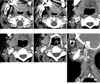
A 54-year-old woman presented with a submental mass for 10 days. She underwent neck computed tomography (CT). The CT scan revealed multiple small lymph nodes in the neck. Incidentally, there was a fenestration of the right IJV (Fig. 1).
Discussion
The IJV is the largest collecting vein of the cranium, the face and the anterior region of the neck (12). Fenestrations or duplications of the IJV are rare variations, but there is an important difference between fenestration and duplication. Downie et al. (10) suggested that the term 'duplication' should be used for those cases where the branches of the anomalous vessel, regardless of artery or vein, remain separate along the whole length of their normal course and that the term 'fenestration' should be used for those cases where branched vessels rejoin a single normal vessel. According to this suggestion, our case is not a duplicated IJV but a fenestrated IJV.
Although fenestration of the IJV is clinically insignificant, preoperative diagnosis of a fenestrated IJV may be important because the accessory spinal nerve may pass through a fenestration of the IJV and resection or injury of these structures can cause considerable morbidity (3). There have also been several reports about an accessory spinal nerve passing through a fenestration of the IJV (345), but such nerves usually pass superficial to the IJV (2). Because our patient didn't need an operation, we were not able to find the anatomical relation between the IJV and the spinal accessory nerve.
The etiology of fenestrated IJVs is still unclear and several hypotheses have been suggested to describe the embryological basis of this fenestration. Among vascular, neural or bony hypotheses, the vascular theory is the most likely and is usually accepted in the literature (23). Embryologically the IJV has a lateral and medial vein of the head and the accessory spinal nerve passes between them. One of them, usually the lateral vein, disappears. However, a secondary venous ring surrounding the accessory spinal nerve may develop, and this may be an important cause of fenestration of the IJV (2).
Precise diagnosis, bearing in mind a fenestrated IJV as a variation, may be helpful for various surgical dissections and radiological procedures in the neck.
 XML Download