

 PDF
PDF ePub
ePub Citation
 Print
Print


Rhabdomyosarcomas chiefly occur in infants and children and they are far less frequent in adults. Histologically, they are classified into three subtypes: embryonal, alveolar and pleomorphic. While the head and neck are the principal locations for childhood rhabdomyosarcoma, head and neck rhabdomyosarcoma is rare in adults (1). The overall survival is worse for adults than for children (2). The major sites of metastases are lung, bone, bone marrow, liver and kidney.
Several investigators have evaluated the utility of 18F-FDG PET or PET/CT for the assessment of childhood rhabdomyosarcoma at various body sites (34567). However, there are no reports in the medical literature regarding the 18F-FDG PET/CT findings of adulthood sinonasal rhabdomyosarcoma.
We report herein two cases of adulthood sinonasal alveolar rhabdomyosarcoma that showed intense hypermetaboloism and disseminated metastases on 18F-FDG PET/CT in a 48-year-old woman and a 68-year-old woman, along with the CT and MRI findings.
Case Reports
Case 1
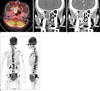
A 68-year-old woman presented with the chief complaints of epistaxis and back pain. Physical examination revealed a polypoid mass at the right middle meatus. The microscopic and immunohistochemical findings of the specimen obtained by endoscopic biopsy of the mass were consistent with rhabdomyosarcoma of the alveolar subtype. The 18F-FDG PET/CT obtained four days after biopsy showed a lobular mass with intense hypermetabolism (SUVmax = 14.6 g/mL) in the right sinonasal area (Fig. 1A). Also noted were diffuse areas of hypermetabolism involving the axial and appendicular skeleton and the lymph nodes of the right retropharyngeal chain and the left level VA, suggestive of disseminated involvement of the bone marrow and lymph nodes (Fig. 1B). This was confirmed by biopsy of the bone marrow and a lymph node of the left level VA. Non-enhanced (NECT) and contrast-enhanced CT (CECT) (Figs. 1C, D) and the gadolinium-enhanced T1-weighted images (Gd-T1WI) showed a large mass with multiple rings of intense enhancement (the "botryoid sign") and bony destruction involving the right nasal cavity and the right ethmoid and sphenoid sinuses. She received 4 cycles of CYVADIC chemotherapy and 5 cycles of chemotherapy consisting of ifosfamide, etoposide and vincristine. She experienced subdural hematoma without a history of trauma 12 months after the initial diagnosis of sinonasal alveolar rhabdomyosarcoma. Sadly, in spite of performing craniotomy with removal of the subdural hematoma, she expired 13 months after the initial diagnosis.
Case 2
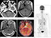
A 48-year-old woman presented with the chief complaints of left nasal obstruction and bloody discharge for several months. Physical examination disclosed a polypoid mass in the left middle meatus. Pre-biopsy NECT demonstrated a large hyperattenuating mass involving the left nasal cavity, the left ethmoid sinus and the bilateral sphenoid sinuses with bony destruction (Fig. 2A). The microscopic and immunohistochemical findings of the specimen obtained by incisional biopsy of the mass were consistent with rhabdomyosarcoma of the alveolar subtype. The Gd-T1WI obtained seven days after biopsy demonstrated a mass with the "botryoid sign" (Figs. 2B, C). 18F-FDG PET/CT performed nine days after the biopsy demonstrated an intensely hypermetabolic mass (SUVmax = 11.9 g/mL) in the sinonasal area (Fig. 2D). Also noted were multiple bone marrow metastases in C3-6, T12 and L3, the sacrum, the pelvic bones, the scapula and the femur (Fig. 2E). She received four cycles of chemotherapy consisting of ifosfamide, etoposide and vincristine. A mass developed in her right breast six months after the initial diagnosis of sinonasal alveolar rhabdomyosarcoma, which was proven to be metastatic rhabdomyosarcoma by ultrasound-guided core-needle biopsy. Unfortunately, she experienced progressively aggravating sepsis and then expired 14 months after the initial diagnosis.
Discussion
Sinonasal rhabdomyosarcoma in adults presents a diagnostic challenge because most benign and malignant sinonasal tumors are seen as solidly enhancing mass on CT or MRI, and they cannot be differentiated from rhabdomyosarcoma. Hagiwara et al. (8) noticed that four of eight cases of rhabdomyosarcoma of the head and neck showed multiple rings of enhancement that resembled a bunch of grapes (the "botryoid sign", on Gd-T1WI. The "botryoid sign" was identified in our two cases on both CECT and Gd-T1WI. The "botryoid sign" is known to be caused by abundant mucoid stroma surrounded by a thin layer of tumor cells (9). Several investigators have reported the SUVmax of rhabdomyosarcoma at various sites of the body (345). Our two cases of adult alveolar rhabdomyosarcoma showed SUVmax of 11.9 g/mL and 14.6 g/mL, respectively, which were far higher than those reported by other studies in which the majority of cases were in the pediatric age group (345).
The initial presentation of alveolar rhabdomyosarcoma with concurrent metastases to the bone marrow and lymph nodes mimicking acute leukemia/lymphoma, has rarely been reported in children (67) or adults (10). Our cases represented sinonasal alveolar rhabdomyosarcoma manifesting intense hypermetabolism of the primary site and disseminated bone marrow metastases at the initial presentation, as demonstrated by 18F-FDG PET/CT. Case 1 presented a diagnostic dilemma because the disseminated metastases involved the bone marrow and cervical lymph nodes at the initial presentation and this mimicked malignant lymphoma/leukemia. Case 2 represented adulthood alveolar rhabdomyosarcoma with breast metastasis, which might represent a sign of disseminated disease and so a poor prognosis. Our review of the literature has yielded two cases of alvelolar rhabdomyosarcoma, each in the orbit and foot, respectively, of two 16-year-old girls, and the tumor showed intense hypermetabolism of the primary sites and disseminated bone marrow metastases (67). We hypothesized that the intense hypermetabolism of the primary site might be associated with disseminated metastases. Our cases also suggest the potential usefulness of 18F-FDG PET/CT not only for the detection of the primary site, but also for the staging of adulthood alveolar rhabdomyosarcoma.
In summary, the diagnosis of adulthood rhabdomyosarcoma can be considered when a sinonasal mass demonstrates the "botryoid sign" on CECT or Gd-T1WI, and intense hypermetabolism and disseminated metastases on 18F-FDG PET/CT. Intense hypermetabolism of the primary site may herald disseminated metastases and thus a poor prognosis. Care must be taken to avoid misdiagnosing sinonasal alveolar rhabdomyosarcoma as lymphoma or leukemia.
 XML Download