

 PDF
PDF ePub
ePub Citation
 Print
Print


Enteroviruses include the coxsackie viruses A and B, poliovirus, echoviruses and enteroviruses 68 to 71. These may be the cause of hand-foot-mouth disease (HFMD) (Coxsackie virus A16, enterovirus 71) (1), herpangina (enterovirus 71), hemorrhagic conjunctivitis (enterovirus 70, Coxsackie virus A24) (2), poliomyelitis (poliovirus), polio-like paralysis or radiculomyelitis (enterovirus 70, enterovirus 71, Coxsackie virus A7 and A24) (34).
The HFMD caused by coxsackie virus A16 or coxsackie virus A10 is usually a mild, self-limiting illness that primarily affects infants and young children and there are usually no central nervous system (CNS) complications (5). Yet enterovirus 71 (EV 71) may cause not only HFMD, but also various neurologic complications such as aseptic or viral meningitis, encephalitis or polio-like paralysis (6). Especially, EV 71 meningitis or encephalitis can occasionally be fatal (7).
The non-contrast enhanced MR imaging findings of EV 71 encephalomyelitis were first published in 1999 by Shen et al. (8). The MR imaging features such as brainstem involvement and an abnormal signal on a T2-weighted image (WI) were described in this article. However, to the best of our knowledge, there have been few reports about the MR features on contrast enhanced T1-WI and the diffusion weighted image (DWI) of EV 71 encephalitis in infants with HFMD.
We report here on the MR imaging findings of a case of acute EV 71 encephalitis that involved the brain stem in a 33-month old girl with HFMD.
Case Report
A 33-month old girl was admitted for general weakness. On the physical examination, she was febrile, anorexia and she had the skin manifestations of erythematous vesicles on both hands, elbows, feet and knees, and ulcers on her lips and tongue. She was diagnosed with HFMD. A neurologic examination revealed drowsiness, lethargy, right lower extremity weakness (grade II) and sensory loss of both lower extremities (right: grade I-II, left: grade III-IV). These neurologic symptoms were suspected to be caused by CNS involvement due to encephalitis, yet she didn't show meningism, cranial nerve (CN) palsies, disturbance of ocular movement or cardiopulmonary complications. The cerebral spinal fluid (CSF) study on the day of admission showed 193 WBCs/uL, a protein level of 25 mg/dL and a glucose level of of 75 mg/dL. The CSF polymerase chain reaction (PCR) corresponding to enterovirus 71 ribonucleic acid (RNA) was positive.
On the day of admission, MR images were obtained with a 1.5T MR machine (Siemens 1.5 T, Sonata, Germany). The T1-weighted spin-echo sequences (the repetition time msec/echo time msec was 486/7.7), the T2-weighted spin-echo sequences (4770/108) and the T1-weighted gadolinium-enhanced spin-echo sequences (425/17.0) were obtained. The gadolinium-enhanced spin-echo sequences were obtained after the administration of 0.2 mL/kg (1 mmol gadolinium per kilogram) gadobutrol (Gadovist; Schering, Weesp, the Netherlands). Diffusion-weighted imaging was performed by obtaining the single-shot spin-echo echo-planar sequences (4500/99) with a b value of 2000 sec/mm2. The section thickness of all the sequences was 5 mm with an intersection gap of 1.0 mm. The brain MR images revealed increased signal intensity (SI) on the T2-WI at the posterior aspect of the medulla, the pontine tegmen, the bilateral dentate nuclei of the cerebellum and the midbrain. DWI also showed increased SI at the posterior aspect of the medulla and pontine tegmen, and there was low SI at the same areas on the apparent diffusion coefficient (ADC) map. She was initially treated with hydration. Three days after admission, the fever was subsided and her mental state was alert. Although she is slowly getting better through rehabilitation for a month, her right lower extremity weakness has not yet fully recovered and she can't stand up without assistance. Yet the sensations of both lower extremities were fully regained.
Discussion
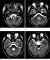
Wu-Chung Shen et al. (8) reported that the typical locations of EV 71 encephalomyelitis were in the posterior aspects of the medulla and pons, the central portion of the midbrain, the bilateral dentate nuclei of the cerebellum and the bilateral ventral horns of the cervical spinal cord. The MR images of our case also revealed high SI on the T2-WI at the posterior aspect of the medulla, the bilateral dentate nuclei of the cerebellum, the pontine tegmen and the midbrain (Fig. 1). However, the reason why EV 71 encephalitis has its characteristic locations of CNS involvement is still unknown.
As we know, there are the dorsal nuclei of CN X, the medial longitudinal fasciculus, the reticular formation and the nuclei of the solitary tract in the posterior aspect of the medulla, the nuclei of CN VI, VII and IX in the posterior aspect of the pons, and the red nuclei, the substantia nigra and the nuclei of CN III and IV in the midbrain. Therefore, EV 71 encephalitis with brainstem involvement can cause various and even serious CNS complications such as lethargy, CN palsies, conjugated disturbance of ocular movement, dyspnea and tachycardia (8). In our case, there were also CNS complications such as drowsiness, lethargy and ataxia due to the acute EV 71 encephalitis. It is important that the locations involved by EV 71 encephalitis should be accurately detected early through the initial MR imaging. This may help predict the cardiopulmonary complications and an improved clinical outcome.
In our case, there was no significant abnormal signal or enhancement on the pre- and post-contrast T1-WI (Fig. 2A) at the areas that showed high SI on T2-WI. This means the lesions were spots of acute inflammation of brain tissue without tissue destruction or the degradation of the blood-brain barrier. If the follow-up MR images obtained later disclose low SI on the T1-WI and high SI on the T2-WI, then the brain tissues will reflect tissue destruction and irreversible damage (8).
In addition to the conventional MR images, the DWI and ADC map were obtained in our case. DWI showed high SI at the posterior aspect of the medulla (Fig. 2B) and the pontine tegmen, and the ADC map revealed low SI at the same areas (Fig. 2C). At the acute stage, these diffusion weighted MR features represent reduced diffusivity and cytotoxic edema. On the other hand, these MR findings on DWI and an ADC map may be associated with clinical outcome like that for herpes simplex encephalitis. Sener RN (9) reported that during the acute stage of herpes simplex encephalitis, the diffusion MR imaging findings that reflect cytotoxic edema may be characterized by severe tissue damage and this is associated with a poor clinical outcome. More cases should be studied on a case by case basis to determine whether or not the diffusion MR imaging findings that reflect cytotoxic edema can be a prognostic factor for EV 71 encephalitis in the acute stage.
In addition to conventional MR imaging, DWI may be helpful for the early detection of acute EV 71 encephalitis in a patient with HFMD.
 XML Download