

 PDF
PDF ePub
ePub Citation
 Print
Print


Calcineurin inhibitors such as cyclosporine and FK506 (tacrolimus) are potent immunosuppressive agents that are widely used in organ transplantation of the kidney. However, these agents can induce neurotoxicity. The spectrum of neurological disturbances caused by calcineurin inhibitors ranges from very mild symptoms such as paresthesia, tremor, headache and flushing to severe changes that may cause a lethal outcome (12). The neurotoxicity of these agents in the central nervous system has been increasingly reported with the widespread use of magnetic resonance imaging (MRI). FK506 leukoencephalopathy has been included within posterior reversible leukoencephalopathy syndrome (PRES), and this has also been caused by cyclosporine, eclampsia, malignant hypertension, renal failure and drug-induced thrombotic thrombocytopenic purpura. The MR finding of FK506-induced PRES are well documented and these include increased signal intensities in both parieto-occipital lobes on the T2-weighted images (T2WI), the diffusion-weighted images (DWI) and the apparent diffusion coefficient (ADC) mapping. These findings indicate vasogenic edema rather than cytotoxic edema (34). However, reports about FK506-induced cerebral infarction are not common.
We report here on a case of acute cerebral infarction after FK506 administration in a kidney transplantation recipient.
Case Report
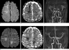
An 11-year-old girl with chronic renal failure due to congenital renal hypoplasia underwent kidney transplantation from her mother. This patient was born prematurily. She had suffered from renal failure for 11 years. After renal transplantation, she was receiving FK506 to prevent graft versus host disease. The dosage of FK506 (approximately 0.2 mg/kg per day) was increased to attain the targeted blood level. The serum concentration of FK506 was maintained between 10-22 ng/mL for 11 post-operative days. Twelve days after renal transplantation, she showed symptoms of left hemiparesis and headache. The serum concentration of FK506 was 10.5 ng/mL and the patient's blood pressure was 160/100 mmHg. Brain MRI was performed. Increased signal intensity on the DWI and decreased signal intensity on the ADC mapping in the right frontal lobe were demonstrated and these findings indicated acute infarction. MR angiography (MRA) showed diffuse stenosis in both the anterior and middle cerebral arteries. Follow-up MRA was obtained one month after the discontinuation of FK506 administration, and the stenosis of both anterior and middle cerebral arteries was improved with a normal caliber of the vessels. The clinical symptoms were also improved.
DISCUSSION
FK506 (tacrolimus) is a macrolide antibiotic and it has immunosuppressant activity similar to that of cyclosporine. However, it is between 10 and 100 times more potent than cyclosporine in terms of its immunosuppressive properties (5). Widespread use of FK506 as an immunosuppressive agent following organ transplantation has led to a number of neurological symptoms. The common clinical features of FK506 neurotoxicity include the sudden onset of seizures, headache, an altered mental status, visual abnormalities, aphasia and hemiparesis. The onset of neurological symptom may be acute or subacute. In solid organ transplant recipients, these major neurological toxicities have been observed in approximately 5% of the patients receiving FK506 and they have occurred at median of 10-13 days after its initial administration (6).
The pathophysiology of both cyclosporine and FK506 neurotoxicity currently remain unclear. Two pathogenic mechanisms have been suggested. One is that the acute hypertension in posterior leukoencephalopathy induces a loss of autoregulation with passive dilatation of the cerebral arterioles; the hydrostatic pressure results in the extravasation of proteins and fluid into the interstitium. Multiple reports of reversible T2 hyperintense white matter lesions with a predominance in the posterior circulation as well as increased perfusion seen on SPECT scanning in patients with posterior leukoencephalopathy support this theory (3). Another theory is vasculopathy. Diffuse vessel injury can lead to cerebral vasoconstriction and related ischemia. FK506-induced leukoencephalopathy was been increasingly reported with the widespread use of the MRI. However, reports about FK506-induced cerebral infarction are rare. We think that the FK506-induced cerebral infarction is caused by vasospasm and this vasospasm may have improved after discontinuing the administration of FK506.
It has been postulated that FK506 neurotoxicity may be similar to hypertensive encephalopathy. However, not all the patients with immunosuppressive induced leukoencephalopathy have hypertension. Hypertensive encephalopathy is the most widely recognized cause of posterior reversible leukoencephalopathy syndrome (PRES), which is a condition that is characterized by the rapid development of vasogenic edema in the posterior head regions (7). Ahn et al. (3) reported the DWI and ADC mapping findings of a FK506 neurotoxicity patient who showed increased signal intensities in both parietooccipital lobes on the T2WI, DWI and ADC mapping. They suggested that vasogenic edema rather than cytotoxic edema may play a pivotal role in the pathogenesis of FK506 neurotoxicity. Kilinc et al. (8) reported that a 18-year-old male with chronic renal failure underwent uncomplicated renal transplantation from his mother using an immunosuppression protocol of mycophenolate mofetil, tacrolimus and prednisolone. Brain MRI on the second day of hospitalization showed signal change in the deep bilateral temporal and occipital regions as well as in the right frontal and right posterior parietal regions. The DWI revealed vasogenic edema in the right frontal, right occipital, and left temporal regions. An autopsy was performed 6 hours after death. The principal pathologic diagnosis was multiple cerebral hemispheric infarctions that were possibly due to vasculitis.
Although reversible vasogenic edema due to cerebrovascular autoregulatory dysfunction is the underlying pathophysiologic mechanism, irreversible lesions resulting from cytotoxic edema can be associated with FK506 neurotoxicity. Kinoshita et al. (4) mentioned that the MR abnormalities related to the FK506 neurotoxicity included in PRES are similar to those observed in hypertensive encephalopathy. However, the cortical laminar necrosis in their other case could have developed after treatment with FK506. The initial images show cytotoxic edema in the gray and white matter. Follow-up studies demonstrate cortical hyperintensity in the boundary zone on the T1-weighted images, and this is consistent with cortical laminar necrosis. They suggested this is probably due to a transient hypoxic-ischemic process. Curro et al. (9) reported the case of a male liver transplant recipient who developed de novo migraine while on FK506 therapy. Brain MRI showed a single focal ischemic lesion in the occipital lobe with no abnormalities in the cortex other than those in the white matter in the remnant brain. MRA suggested vasospasm, which was supported by the findings of a reduction of the lumina of the left anterior and middle cerebral arteries with normal contralateral arteries. They suggest that the combination of rizatriptan and tacrolimus can potentially lead to an excessive risk of clinically evident cerebral vasospasm.
In conclusion, we report here on a case of cerebral acute infarction caused by vasospasm in a patient with FK506 neurotoxicity. On DWI, there were areas with increased signal intensity in the cerebral hemispheres. The areas showing increased signal intensity on DWI and T2WI demonstrated decreased signal intensity on the ADC map image. Follow-up MRA was obtained after the discontinuation of FK506, and the stenosis of both anterior and middle cerebral arteries was improved with a normal caliber of the vessels. These findings suggest that cytotoxic edema is the main cause of the increased signal intensities seen on T2WI, and vasospasm-induced infarction rather than breakdown of autoregulation played a pivotal role in the pathogenesis of FK506 neurotoxicity. Discontinuing FK506 administration may result in improvement of the vasospasm.
 XML Download