

 PDF
PDF ePub
ePub Citation
 Print
Print


Breast reconstruction with TRAM flaps has become an increasingly common surgical procedure among breast cancer patients who undergo a mastectomy (1). TRAM flap reconstruction involves the transfer of lower abdominal skin and subcutaneous fat to the chest defect at the mastectomy site (2). In this procedure, the epidermal inclusion cyst can be developed at the surgical site if the de-epithelized portion of buried skin flaps retains the epithelial elements. This epidermal inclusion cyst, which can be mistaken for a recurrent cancer, may lead to a false positive finding on a mammogram.
To our knowledge, reports on epidermal inclusion cysts after breast reconstruction surgery have not been previously described in the literature. So, we report our experience of an epidermal inclusion cyst in a patient who underwent a total mastectomy with breast reconstruction and TRAM flaps.
Case Report
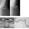
A 50-year-old woman was admitted to our hospital for a follow-up mammography. She underwent a left total mastectomy and reconstruction with TRAM flaps for treatment of the ductal carcinoma in situ, about 6-months prior. The mammography showed findings suggesting that the postoperative change and an oval-shaped, circumscribed partially indistinct marginal isodense mass at the 2 o'clock position of the left breast (Fig. 1A). The mass was believed to be benign, such as fat necrosis or benign granuloma, so the patient was recommended to receive follow-up mammography 6-months later.
The 6 month follow-up showed an increase in the size of the previously detected mass at the 2 o'clock position from about 10 mm to 13 mm in diameter (Fig. 1B). On ultrasonography, the lesion was located in the peripheral portion of reconstructed breast, adjacent to the incision site and subcutaneous fat layer. The mass showed predominantly hypoechoic with heterogeneous internal echoes and circumscribed margin (Fig. 1C). The Power Doppler did not show any vascularity. The sonographic appearance was believed to be benign. However, since the size of the mass increased, it was categorized as BI-RADS 4a and an ultrasound-guided 14-gause core needle biopsy was undertaken. The histology specimens showed a cyst which was lined by stratified squamous epithelium and filled with keratin flakes (Fig. 1D).
The follow-up mammography of the left breast obtained at 4 months after a needle biopsy showed no change in the appearance of the mass.
Discussion
An epidermal inclusion cyst is a benign cutaneous or subcutaneous lesion that is lined with mature stratified squamous epithelium (3). An epidermal inclusion cyst of the breast is rare and only a few cases have been reported in the literature (4).
Epidermal inclusion cysts in the breast are believed to arise by a few different mechanisms (56); most commonly from inflammatory downward growth of epithelium by an inflammatory response of obstructed hair follicles. Second, the trauma itself may stimulate epithelial proliferation and create the cysts. Third, they can be result of squamous metaplastic transformation of the usual columnar cells of the breast, and finally, they might be congenital, arising from cell nests remaining from the embryonal mammary ridge.
An epidermal inclusion cyst in the breast may also be associated with breast surgery. The formation of an epidermal inclusion cyst after a reduction mammoplasty has been reported (7). The breast reduction surgery generally includes reducing the dermal envelope of the breast, resecting parenchyma, and elevating the nipple-areolar unit on a vascularized tissue pedicle of mammary tissue. If epithelial elements are retained within the infolded dermoglandular structures or at any other skin closure, epithelial inclusion cyst formation may occur (7).
In our patient's case, the TRAM flap reconstruction technique also included the procedure of the de-epithelizing the portion of the buried skin flaps. So, the epidermal inclusion cyst may have developed by epithelial elements which are retained on the skin flap, which are similar mechanisms to a reduction mammoplasty.
At mammography and ultrasound, epidermal inclusion cysts may appear with typical benign features; as well-defined, cystic, or solid mass (8). In our patient's case, she had undergone a left mastectomy due to breast carcinoma that causes more difficulty in assessing a newly developed benign looking mass after reconstructive surgery. In addition, the size of the mass increased, so it was categorized as BI-RADS 4a and an ultrasound-guided core needle biopsy was performed.
We presume there is one clue indicating the presence of an epidermal inclusion cyst after a mastectomy with reconstructive surgery for breast cancer. That is the location of the lesion. By using the TRAM flap technique, the epidermal inclusion cyst may develop just below the incision site, because that is the only de-epithelized portion of reconstructed breast. So, we can expect the possibility of an epidermal inclusion cyst when the mass is located in the peripheral portion of the reconstructed breast, adjacent to the incisional site. Correspondingly, in our patient's case, the epidermal inclusion cyst was located in the peripheral portion of reconstructed breast, adjacent to the operative scar.
An epidermal inclusion cyst is usually located in the dermis; however, some have also been reported in deeper locations within the breast parenchyma, with or without previous breast surgery (479). In our patient, the cyst has developed within the deep portion of the subcutaneous fat layer instead of the breast parenchyma, because she underwent a total mastectomy with breast reconstruction and had no remaining breast parenchyma. Also, there is no direct connection to the skin surface with an epidermal inclusion cyst. Therefore, we can suspect another possibility of the epidermal inclusion cyst when the mass is located in a deep portion of the subcutaneous fat layer of the reconstructed breast, without connection to the skin surface.
There are many reports about the usefulness of needle or fine needle aspiration for the diagnosis of epidermal inclusion cysts (710). Also, in our patient's case, our pathologist thought the results of core-needle biopsy were considered diagnostic to avoid surgical biopsy.
In conclusion, an epidermal inclusion cyst within the breast is rare benign lesion, but occasionally that need to differente it with a malignant lesion, especially in the case of post-operative breast cancer. The mammographic and sonographic features may not offer the confident diagnosis of an epidermal inclusion cyst, so the diagnostic needle or excisional biopsy is essential. However, there are some possibilities to diagnose an epidermal inclusion cyst when the lesion is located in the peripheral portion of reconstructed breast, adjacent to the incision site and a deep region within the subcutaneous fat layer.
 XML Download