

 PDF
PDF ePub
ePub Citation
 Print
Print


Myositis ossificans is a benign, self-limiting, non-neoplastic formation of heterotopic bone in the skeletal muscle (12345). Although myositis ossificans can occur anywhere in the body following trauma, it is predominantly localized to high-risk sites of injury, such as large muscles of the extremities (234). Most lesions are related to direct major trauma or repeated minor injuries. No history of trauma is present in approximately 40% of patients (23).
In the English medical literature, there are many reports on myositis ossificans that included radiographic, CT and MR imaging findings (1234567). The CT and MR imaging findings of myositis ossificans involving the psoas muscle have been reported twice (27).
We could not identify any reports on CT and MR imaging findings of the psoas muscle following lumbar spine fracture in the radiological literature. We report the CT and MR imaging findings of a 64-year-old man who developed myositis ossificans of the psoas muscle following lumbar spine fracture.
Case Report
A 64-year-old man presented with a 5-day history of lower back pain after a fall. On admission, his vital signs were normal. Laboratory values, including C-reactive protein and erythrocyte sedimentation rate, were within the normal limits.
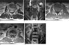
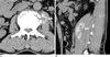
Plain radiography of the lumbar spine revealed compression fracture in L3. MRI examination was performed on a 1.5T MR unit (Siemens, Erlangen, Germany). Spin-echo T1- and turbo spin-echo T2-weighted MR images demonstrated L3 compression fracture and a left psoas mass on the fifth post-trauma day. The mass was of high signal intensity with no peripheral hypointense rim on turbo spin-echo T2-weighted images (Fig. 1A, B). The mass was isointense on spin-echo T1-weighted image, compared to the muscle (Fig. 1C). Gadolinium-enhanced T1-weighted images showed diffuse homogenous enhancement of the mass without central necrosis (Fig. 1D, E). A pre-contrast CT scan was taken in the third post-trauma week showed a well defined ossified peripheral rim and a central non-ossified area of low attenuation (Fig. 2A, B).
Myositis ossificans was diagnosed based on these CT findings. We chose to manage the patient conservatively and avoided invasive diagnostic and treatment procedures.
Discussion
Myositis ossificans is a benign, non-neoplastic heterotopic formation of bone within the muscle or soft tissues that usually develops in adolescents and young adults, typically following trauma (1234). Although myositis ossificans can occur almost anywhere in the body, it commonly affects large muscles of the extremities, such as the thigh, buttocks, elbow and less often, the shoulder and calf (234). Although the exact etiology remains unknown, the pathophysiology of myositis ossificans is thought to be a post-traumatic inflammatory process within the skeletal muscle (2).
Typical clinical presentation of this condition includes rapid onset of pain, palpable mass, flexion contracture, local heat and swelling as well as decreased range of motion (3). In our patient, the only presenting symptom was lower back pain.
Histologically, myositis ossificans tends to display a typical zonal pattern. At 6-8 weeks, a lacy pattern of new bone formation in the peripheral zone is formed, and the zonal pattern of peripheral maturation is the most important diagnostic feature. Histologically, the central zone is comprised of undifferentiated mesenchymal cells with high-grade mitotic activity, which may suggest malignant neoplasm. Therefore, early biopsy of the center lesion may lead to mis-diagnosis and clinicians should exercise caution (36).
The radiologic findings of myositis ossificans mirror the histologic pattern of maturation. The earliest radiologic change appears within 1 to 2 weeks as a soft tissue mass, which may be accompanied by faint periosteal new bone formation. By 3-4 weeks, floccular calcification appears in the soft tissue mass. By 6-8 weeks, a lacy pattern of new bone formation in the peripheral zone is formed. By 5 to 6 months, the mass shrinks and maturation is completed (3).
The cross-sectional CT and MRI findings of myositis ossificans in the English medical literature are well known (1234567). Previously published CT findings of myositis ossificans included a well-defined ossified peripheral rim associated with a central non-ossified area of low density after 4-6 weeks that mimicked the histologic zonal architecture. CT has been the gold standard in characterizing the typical pattern of myositis ossificans (23). In our case, a pre-contrast CT scan was taken in the third post-trauma week which demonstrated a well-defined diagnostic ossified peripheral rim in the psoas muscle.
The MRI appearance of myositis ossificans correlates with the stage of maturation and the histologic pattern of the lesion (13). In the early stages, T2-weighted images may show an in-homogenous focal mass with high central signal intensity. As the lesion matures and the peripheral ossification becomes denser, the images show a hyper-intense center surrounded by a hypo-intense rim corresponding to peripheral ossification (3). In our patient, MRI of the lumbar spine was performed on fifth post-trauma day. It showed high signal intensity with no peripheral hypointense rim, and the appearance appeared to be non-specific. As such, the MRI findings of myositis ossificans often lead to erroneous diagnosis of sarcoma or infectious myositis (23). In our patient, no clinical or laboratory evidence of infection or tumor was present, and concomitant fracture of L3 along with typical patterns of myositis ossificans on CT excluded sarcoma and infectious myositis. Sirvanci et al. (2) reported the CT and MR imaging findings of myositis ossificans of the psoas muscle, but a history of trauma was not apparent in that case. Kim et al. (7) reported the CT and MR imaging findings of myositis ossificans of the psoas muscle following lumbar compression fracture, and the diagnosis was confirmed with CT-guided biopsy. In our patient, the diagnosis of myositis ossificans was made based on typical CT imaging findings of myositis ossificans. After discussions with other clinicians, we chose not to perform CT-guided biopsy and focused on managing the patient conservatively.
In conclusion, myositis ossificans should be considered a differential diagnosis when a well-defined mass with homogeneous enhancement is seen within the psoas muscle following fracture of the lumbar spine, particularly when tumor and infection have been excluded by investigations.
 XML Download