

 PDF
PDF ePub
ePub Citation
 Print
Print


Hemoperitoneum is an emergency life-threatening condition that is caused by blunt abdominal trauma, and the spleen and liver seem to be the most frequently injured organs (123). Splenic enlargement is often observed on follow-up computed tomography (CT) examination after traumatic hemoperitoneum, but this splenic enlargement is known to return to normal size after physiologic contraction of the spleen rather than being true splenic enlargement (45). The spleen serves as a reservoir for formed blood elements and it is known that red blood cells are released into the circulation by splenic contraction in the face of bleeding (567).
Goodman and Aprahamian (4) described an average 25% increase of splenic volume on follow-up CT examination after blunt abdominal trauma, which they interpreted as being initial physiologic splenic contraction with an eventual return to normal size. They emphasized that this enlargement of the spleen on follow-up CT should not be considered an indicator of a deteriorating condition. Although they considered the volume of the spleen on follow-up CT examination as normal, it is difficult to know the normal volume of the individual spleen in the absence of performing a CT examination before the traumatic event.
We recently found that the splenic volume was decreased on long-term follow-up CT examination, as compared with that seen on the previous follow-up CT, in a patient with a history of traumatic hemoperitoneum. This experience alerted us that the spleen might enlarge rather than return to normal size. We retrospectively studied the serial changes of splenic volume in patients with traumatic hemoperitoneum and who underwent long-term follow-up CT examination. We presumed that the splenic volume on the long-term follow-up CT examination might be close to the pretraumatic splenic volume. To the best of our knowledge, no study has yet focused on the long-term follow-up study of the splenic volume changes in patients with hemoperitoneum.
The purpose of this study is to evaluate the initial and early changes of splenic volume in patients with traumatic hemoperitoneum, based on the estimated normal splenic volume obtained on the long-term follow-up CT examination.
Materials And Methods
Study Group
All the consecutive patients who underwent abdominal CT examinations for traumatic intraperitoneal hemorrhage after blunt abdominal trauma at our institution between the years 2004 to 2008 were retrospectively reviewed to determine their eligibility for this study. Patients were included in this study if they had undergone an initial CT within one day after trauma, at least one follow-up CT within 30 days and at least one additional follow-up CT obtained 30 days after trauma. Patients with splenic injury, infectious disease, chronic liver disease, hematologic disorders or other underlying disease that cause splenomegaly were excluded. The study group consisted of 20 patients (18 men and two women, age range: 9-68 years, average age: 42.0 years). A review of the medical records was conducted to calculate the total amount of blood transfusion. Approval from our institutional review board was obtained for this retrospective study.
CT Examination
CT examinations were performed on a single-detector helical CT (HighSpeed, GE Healthcare), a 4-multidetector CT (MDCT) (LightSpeed plus, GE Healthcare) or a 64-MDCT scanner (LightSpeed VCT, GE Healthcare) for generating axial images with contiguous 5-mm-thick sections.
A total of 101 CT examinations were performed for the 20 patients. The average was 5.1 CT examinations per patient (range: 3-11). The CT examinations were divided into three groups according to time interval: for the initial CT, CT examinations performed within 24 hours after trauma; for the early follow-up CT (early CT), CT examinations were performed within 30 days after trauma; for the late follow-up CT (late CT), CT examinations were performed 30 days after trauma. The number of initial CT examinations was 20, the number of early CT examinations was 43 (average: 2.2, range: 1-4) and the number of late CT examinations was 38 (average: 1.9, range: 1-8).
Image Analysis
Two radiologists retrospectively reviewed all the CT images on a PACS workstation (Radpia, Hyundai Information Technology). They evaluated the presence of intraperitoneal hemorrhage and visceral organ injuries by working in consensus. One radiologist used an area-measuring tool and the summation-of-areas technique on the CT scans to measure the volumes of the spleen at the maximal magnification of a PACS monitor. The cross-sectional area of each image of the spleen was determined on the monitor by tracking the margin of the spleen with a built-in cursor controlled by a mouse. The section volumes were then calculated by multiplying each cross-sectional area by the section thickness and these were added to determine the total splenic volume. The validity and reproducibility of this method have been previously reported (8910). If two or more early CT examinations were performed, then the largest splenic volume was recorded, and if two or more late CT examinations were obtained, the splenic volume of the last CT examination was then recorded. The average time interval between the traumatic event and the last CT examination was 243.9 days (range: 31-980 days).
On the presumption that the volume of the spleen on the last CT was close to the normal volume, the relative splenic volume on the initial CT (initial RSV) was calculated as follows: [(volume on the initial CT) × 100]/volume on the last CT]. The relative splenic volume on the early CT (the early RSV) was also calculated as follows: [(volume on the early CT) × 100]/volume on the last CT]. The relative splenic volume with less than a 10% increase or decrease was considered to have no significant change.
According to the initial RSV, the patients were divided into two groups: the splenic contraction group (less than 90% of the initial RSV) and the normal group. According to the early RSV, the patients were also divided into two other groups: the splenomegaly group (more than 110% of the early RSV) and the normal group. We further divided the patients into four groups based on the combination of the initial and early RSV for analyzing the pattern of serial changes of the splenic volume: group A (initial splenic contraction and early splenomegaly), group B (initial splenic contraction and a normal early RSV), group C (a normal initial RSV and early splenomegaly) and group D (normal initial and early RSV).
For a more meaningful estimate of the amount of intraperitoneal fluid, we used a hemoperitoneum score for the initial CT examination. The collections of intraperitoneal fluid were categorized into six sites: 1) the perihepatic space, 2) the perisplenic space, 3) the right paracolic gutter with including the hepatorenal fossa, 4) the left paracolic gutter, 5) the mesentery and 6) the pelvis including the pouch of Douglas. Two radiologists working in consensus selected the one representative image of the largest amount of intraperitoneal hemorrhage for each of the six sites. The dimension of the intraperitoneal fluid on each representative image was determined at maximal magnification on a PACS monitor by one radiologist using an area-measuring tool. The total size of the intraperitoneal fluid in a patient was calculated as the sum of the size of the intraperitoneal fluid seen on each image. To complete the hemoperitoneum score, one point was given for a 1,000 mm2 total size of the intraperitoneal fluid.
Statistical Analysis
Statistical analysis was performed using SPSS version 14.0 for Windows. A paired t-test was performed to evaluate the statistical differences on comparing the splenic volumes of the initial and early follow-up CT examinations. We also determined the correlations between the initial RSV and the hemoperitoneum score and between the early RSV and the volume of blood transfusion by using Pearson correlation coefficients. P values <0.05 were deemed significant.
Results
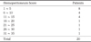
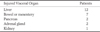
The average hemoperitoneum score of all the patients was 9.9 (range: 1-39) and a relatively large amount of hemoperitoneum (hemoperitoneum score > 10) was seen in 8 patients (40.0%) (Table 1). Visceral injuries were seen in all the patients and the liver was the most frequently injured organ (12 patients, 60%) (Table 2). Transfusions were necessary for 18 patients (90%), and four patients (20.0%) required surgical procedures.
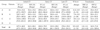
The average RSVs of the initial and early CT were 62.0% and 133.3%, respectively, and all the patients showed an increase of the splenic and relative splenic volumes on the early CT as compared with that of the initial CT (p=0.000) (Table 3). Initial splenic contraction (groups A and B) was seen in 18 patients (90.0%) and the average initial RSV of the splenic contraction group was 58.1%. There was no significant correlation between the initial RSV and the hemoperitoneum score (p=0.529).
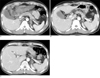
Early splenomegaly (groups A and C) was seen in 14 patients (70.0%) and the average early RSV of the splenomegaly group was 146.5%. The change in the early RSV peaked 13.8 days after trauma. There was no significant correlation between the early RSV and the amount of transfusion (p=0.514). Patients with initial splenic contraction and early splenomegaly (group A) were most common (12 patient, 60.0%) (Fig. 1).
Discussion
The spleen is the largest lymphoid organ in the human body and it serves numerous functions such as formation of antibodies, production of lymphocytes and monocytes, filtration, phagocytosis and destruction of red blood cells (56). It also serves as a storage site for formed blood elements (56). Although the spleen contains a small volume of red blood cells, this small number of red cells can be effectively infused into the circulation by splenic contraction in the face of acute physiological stress, including hemorrhage, exercise, diving, breath holding and drowning (567). This splenic contraction is controlled by the actions of postganglionic fibers on the capsule and trabeculae (5). Our study revealed no significant correlation between the initial RSV and the hemoperitoneum score. This means initial splenic contraction is related to the systemic effect of trauma rather than to only the effect of bleeding. The effect of the total severity of trauma was not included in our study.
Goodman and Aprahamian (4) reported that the average splenic volume increased 25% between the initial and follow-up CT examinations in the patients with blunt abdominal trauma and 57% of their patients had an average increase in splenic volume of 56%. This splenic enlargement was believed to represent a return to normal size after physiologic contraction. But in their study, the time interval between the initial and follow-up CT examinations was less than 7 days and there was no long-term follow-up study. Our study also revealed a significant increase of the splenic volume between the initial and early follow-CT examinations, but the early RSV was significantly increased in 14 patients (70%), with the average early RSV being 146.5%. This result means there was early splenomegaly rather than a return to normal size after the initial splenic contraction. Initial physiologic splenic contraction with a return to the normal early RSV was seen in 6 patients (30%). Our study revealed that the most common pattern of the serial changes of splenic volume was initial splenic contraction, early splenomegaly and a late return to normal size (group A), which was all seen in 12 of the patients (60%). The early RSV peaked 13.8 days after trauma in our study. But Goodman and Aprahamian (4) reported that the changes in splenic volume peaked 6 days after trauma, and this difference is thought to be related to the relatively short follow-up interval of their study.
The causes of diffuse splenomegaly are diverse and they can be categorized on the basis of pathogenesis: 1) congestive splenomegaly that includes portal hypertension and infectious or inflammatory diseases, 2) hyperplastic splenomegaly that includes hemolytic anemia, 3) infiltrative splenomegaly and 4) splenic neoplasm (511). The physiology of early splenomegaly after intraperitoneal hemorrhage may be multifactorial. Transfusion can be considered as one cause of splenomegaly (4). The spleen is composed of two functionally and morphologically distinct compartments, the red pulp and the white pulp, and the red pulp is a blood filter that removes foreign material and damaged erythrocytes (1213). Hyperplastic splenomegaly is caused by hypertrophy resulting from the removal of circulating abnormal blood cells (4). Yet we did not find significant correlation between early RSV and the amount of blood transfusion. The second pertinent fact is that the circulating blood volume can influence the volume of the spleen (41314). With aggressive fluid replacement, the spleen may enlarge in response to the increased circulatory volume. In our study, it was difficult to calculate the total volume of a patient's fluid replacement and intake. Last, the spleen also may enlarge due to increased portal vein resistance in patient with liver injury.
Early splenomegaly after hemoperitoneum has not been previously reported and its clinical significance is not yet established. Goodman and Aprahamian (4) reported that splenic enlargement after blunt abdominal trauma was not an indicator of clinical deterioration and this represented a return to normal size rather than true enlargement. But our study revealed that true splenic enlargement after hemoperitoneum commonly occurred. This splenic enlargement was not necessarily a sign of clinical deterioration, but rather, it was a sign of circulating volume overload.
Several limitations of our study should be mentioned. First, the time interval of the early and late follow-up CT examinations was heterogeneous. Second, many patients suffered from multiple injuries, including extraabdominal damage. Our study did not include the effect of the total severity of traumatic injury. Last, our study was not designed to consider the influence of other factors such as age, gender, operation and the total volume replacement. An operation for hemoperitoneum may be another factor of physiologic stress. We could not compare the degree of the splenic volume changes between the group that received an operation or conservative treatment due to the small case numbers.
In conclusion, physiologic splenic contraction was seen in most of the patients who had hemoperitoneum, and thereafter early splenomegaly was commonly seen before a return to normal size. Early splenomegaly may be related to blood transfusion, a circulating volume overload or increased portal vein resistance with liver injury.
 XML Download