

 PDF
PDF ePub
ePub Citation
 Print
Print


Giant cell tumor (GCT) is a benign neoplasm that usually involves the metaphysis and epiphysis of long bones. Although the spine is the fourth leading location of GCT, most of these lesions occur in the sacrum, followed by the thoracic, cervical and lumbar vertebrae (1). GCT of bone is a benign, but potentially aggressive lesion that shows local recurrence and metastases. Metastatic lung disease rarely originates from a benign tumor, but this can occur from GCT of bone (2). The incidence of lung metastases from a histologically proven GCT has range from 1% to 9% (3). To the best of our knowledge, this is the first reported case of a thoracic spinal GCT that simulated a mediastinal mass, along with dispaying benign pulmonary metastases, in the medical literature. Here we present a case of a histologically proven thoracic spinal GCT that presented as a mediastinal tumor with pulmonary metastases.
Case Report
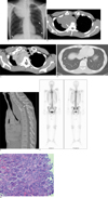
A 29-years-old man was admitted to the hospital for evaluation of active pulmonary tuberculosis. He had been in close contact with a person who was recently diagnosed with active pulmonary tuberculosis. The patient had developed a mild cough and back pain, but there were no other respiratory symptoms. The physical examination and the results of the laboratory tests, including examination for acid-fast bacilli in sputum smears, were unremarkable. Chest radiography showed a huge soft-tissue mass in the right upper mediastinum (Fig. 1A). Moreover, several sharply marginated, round nodules were observed in both lungs (Fig. 1A). The upper mediastinal structures such as the trachea and the aortic arch were not displaced by the mass. We could not find any bony abnormality in the thoracic spine. Computed tomography (CT) showed a large mass with marginal thick shell-like calcification at the right paravertebral space from the T1 to T6 vertebral bodies (Fig. 1B). The paraspinal mass was surrounding the vertebral bodies and a small soft tissue mass also existed in the left paraspinal region. On contrast enhanced CT, the paraspinal mass lesion showed highly enhancing features and some low-attenuation features, and an enhancing soft tissue lesion was also detected at the left epidural space in the spinal canal (Fig. 1C). Multiple small, sharply marginated, round nodules were found to be scattered in both lungs (Fig. 1D) and the sagittally reformatted images showed the total collapse of the T3 vertebral body (Fig. 1E). We believed it was not easy to recognize the bony abnormality on the chest PA film because the T3 vertebral body was totally collapsed. The skeletal scintigram with Tc-99m Hydroxymethylene Diphosphonate showed uneven intense hot uptake at the T3 and T4 vertebral bodies (Fig. 1F). The patient refused further imaging studies such as magnetic resonance imaging (MRI) or positron emission tomography/computed tomography (PET/CT) owing to claustrophobia.
The patient underwent incisional biopsy for the paraspinal soft-tissue mass and wedge resection for the lung nodule. The final pathologic diagnosis was GCT with benign pulmonary metastasis (Fig. 1G).
Discussion
Giant cell tumor represents approximately 5% of all bone tumors and over 20% of all benign bone neoplasm. Giant cell tumors usually involve the metaphysis and epiphysis of long bones. Although giant cell tumors are benign, they have an aggressive tendency to recur locally and show local invasion, and they have low potential for distant metastasis.
Although the spine is the fourth leading location of GCT, most of these lesions occur in the sacrum, and only 2% to 5% of these tumors are found in the vertebrae above the sacrum (1). Sanjay et al. (1) documented only 24 cases of giant cell tumor of the spine at the Mayo Clinic over a 34-year period from 1955 to 1989.
Spinal giant cell tumors are usually an expansile lesion with bone destruction that affects the vertebral body. These features are different from other spinal tumors that mainly affect the posterior elements such as aneurismal bone cyst, osteoid osteoma and osteoblastoma (4). Because extension to the paraspinal soft tissue is often apparent, thoracic spinal giant cell tumor could simulate posterior mediastinal neoplasm (5). Among the posterior mediastinal lesions, the pathologic conditions that can manifest as a paraspinal soft tissue mass include neurogenic tumor, adenopathy, infectious spondylitis, spinal trauma, spinal tumor and extramedullary hematopoiesis. Except for primary spinal tumor, the other posterior mediastinal lesions may not destroy a vertebral body, as happened in this case, and they are not a large paraspinal soft tissue mass.
The radiographic characteristics of spinal giant cell tumors are considered to be a round or oval extrapleural mass with shell-like calcification of the marginal lesions and any mineralized matrix is absent. Extension into the posterior elements and paraspinal soft tissues and associated vertebral collapse are often apparent (6).
On CT scans, a spinal giant cell tumor has been reported to show a homogeneous hypervascular appearance with contrast enhancement (4). Some degree of heterogeneity can be observed because of hemorrhage or necrosis in the tumor (4).
MR images provide more information on both the tumor location and the possible extension of the tumor. The MR images of a giant cell tumor generally show low to intermediate signal intensity on the T1-weighted MR images. On the T2-weighted images, giant cell tumors often have low to intermediate signal intensity caused by the relatively high collagen content of their fibrous components and the hemosiderin within the tumor (7).
As state above, giant cell tumors are benign neoplasm, but they are locally aggressive. Ronald et al. reported that the atypical imaging features of benign giant cell tumors are a compression fracture, posterior subluxation and involvement of the intervertebral disc space (8). Because of these features, giant cell tumors can mimic malignant lesions such as chordoma (8). Further, when a spinal tumor is locally aggressive, giant cell tumor should not be excluded from the differential diagnosis. In our case, complete obliteration of the T3 vertebral body was noted and involvement of the intervertebral disc couldn't be evaluated because the patient refused to undergo MRI.
Sakurai et al. (5) reported the radiologic imaging results in a case of GCT of the thoracic spine and that simulated mediastinal neoplasm, and these imaging results were similar to our case, except for the presence of bilateral pulmonary metastases. Compared with Sakurai's case and the reported typical radiographic appearances, our case also show characteristic radiographic features, including an extrapleural hypervascular mass with some degree of heterogeneity and marginal shell-like calcification.
Lung metastasis from benign giant cell tumor was first reported by Finch and Gleave in 1926 (2). The risk of metastases from a benign giant cell tumor is between 1.8% and 9.1% (3). Beside the lung, rare cases of metastases to other sites have been reported: the lymph nodes, liver, soft tissue, brain, mediastinum, scalp, kidney and penis. The bilateral pulmonary metastases played an important role for us to misdiagnose this GCT as a malignant bone tumor.
GCTs exhibit a varied appearance on bone scintigraphy and they may have a diffuse or homogeneous uptake pattern or "doughnut" or "incomplete doughnut" patterns (9). The "doughnut" sign on a bone scan is nonspecific and this may arise secondary to central necrosis and the surrounding hyperemia (tumor or abscess), or this may be related to a peripheral reparative bone reaction found within active infection, trauma or neoplasm. This pattern has been reported in simple bone cyst, eosinophilic granuloma, aneurysmal bone cyst and bone infarction. The presence of a secondary aneurysmal bone cyst within a GCT has been suggested as to be explanation for the "doughnut" pattern seen on scintigraphy (9). In our case, uneven intense uptake was noted on bone scintigraphy.
Although a thoracic spinal GCT is a benign neoplasm, it can easily simulate a malignant neoplasm because of its locally aggressive features, such as compression fracture and the formation of a large paraspinal soft tissue mass. Moreover, GCT has an ability to metastasize, so when locally aggressive spinal lesion is combined with pulmonary metastases, this is very difficult to diagnose correctly. In conclusion, we report here on a case of thoracic spinal GCT that presented as a huge mediastinal mass and pulmonary metastases.
 XML Download