

 PDF
PDF ePub
ePub Citation
 Print
Print


The number of patients with end stage renal disease requiring catheter-based hemodialysis has increased in recent decades. Catheter-based hemodialysis is commonly used during the time it takes for arteriovenous fistula maturation. Conventional venous access sites such as the subclavian vein (SCV) and internal jugular vein (IJV) had been used for catheter placement. However, the SCV should be preserved from catheterization, with consideration of the risk of venous stenosis or thrombosis as complications after a procedure in a patient where fistula or graft creation is planned. The rate of complications for an SCV approach is higher than for an IJV approach. The IJV has become the preferred primary venous access site for hemodialysis catheter placement with real-time ultrasound guidance, and as a result, can reduce the risk of procedure-related complications (1234).
Recently, several studies have reported that the external jugular vein (EJV) could be used as an alternative venous route for catheter placement with an acceptable technical success rate and low complication rate when the use of the IJV was not feasible (567). However, no report has evaluated the EJV as a primary venous access site when the use of the IJV is feasible. The purpose of this study was to evaluate the feasibility that a dilated EJV can be a primary venous access site for tunneled hemodialysis catheter placement instead of feasible IJV.
Materials and Methods
We performed the placement of a tunneled hemodialysis catheter in 173 patients from January 2008 to April 2009. Among the patients, we attempted to place a large bore tunneled hemodialysis catheter through a dilated right EJV in 42 patients (28 men, 14 women; age range, 22-86 years; mean age, 59 years). Causes of chronic renal failure included hypertension in nine patients, diabetes mellitus in 20 patients, immunoglobulinin A nephropathy in five patients and of unknown origin in eight patients.
Procedure
An interventional radiologist placed all catheters. Antibiotic prophylaxis were not routinely administered for catheter placement. All patients received an intravenous injection of pethidine-HCl (Demerol) before a procedure to control for pain. The neck was examined by ultrasound (EnVisor HD; Philips, Bothell, WA USA) using a 5-12 MHz linear phased array transducer prior to the procedure. The course and caliber of the EJV and IJV were also evaluated by ultrasound. The diameter of the IJV was large enough to accommodate the catheter in 34 patients and small (≦5 mm) in eight patients. The neck and upper chest were prepared with a standard sterile technique. Local anesthesia was achieved with a 2% lidocaine injection. The EJV approach was tried as a primary venous access route for hemodialysis catheter placement if the dilated EJV diameter was greater than 5 mm. If the dilated EJV diameter was smaller than 5 mm, we used the IJV since it is difficult to access a non-dilated EJV. The right EJV was punctured above the clavicle using a 22G angiocath (BD Angiocath plus, Boin Medica, Kyungbuk, Korea) (n=33) or a 21 G micropuncture needle (Cook, Bloomington, IN, USA) (n=9) under real-time ultrasound guidance in all patients. After the EJV was successfully accessed, a 0.018-inch guidewire (Cook, Bloomington, IN USA) from a micropuncture set was advanced into the superior vena cava, and then a 4 F micropuncture exchange dilator was inserted over the wire. Venograms were obtained for evaluation of the course of the vein and patency of the central venous system in one case of failure of the peel away sheath insertion. The guidewire was exchanged with a stiff 0.038-inch wire from a hemodialysis catheter set and the wire was advanced into the right atrium or inferior vena cava. The venotomy site was dilated with a 12 F dilator, and then a 15 F peel-away sheath was inserted carefully over the guidewire under fluoroscopic guidance to ensure that the guidewire did not kink or cause angulation.
A subcutaneous tunnel that extended from the incision to a point 7 cm to 9 cm above the lateral portion of the chest wall was made and the catheter was inserted through the upper portion of the subcutaneous tunnel with a Dacron cuff placed 1 cm to 2 cm from the lower end of the tunnel. Tunneled hemodialysis catheters (14.5 F Soft-Cell chronic dual-lumen catheters; Bard Access Systems, Salt Lake City, UT, USA) with lengths of 19 cm (n=36) or 24 cm (n=6) were used according to the length that was measured using guidewire from the incision site to the atriocaval junction as determined by fluoroscopy.
We performed catheter insertion through the peel-away sheath using finger-pinch technique during expiration. The catheter tips were positioned at the atriocaval junction or in the right atrium. After catheter placement, heparin (100 IU/mL heparin sodium; Green Cross, Seoul, Korea) was injected into each lumen of the catheter for prevention of thrombosis. At the time of insertion, we recorded the procedure time, defined as the time that had elapsed from local anesthesia to the final suture, as well as any problems or complications associated with catheter insertion.
Follow-up
Technical success was defined as catheter introduction into the venous system with the tip positioned in the desired location, and with adequate catheter function in the hemodialysis room. A rate of 300 mL/min was considered as an adequate rate of blood flow in adult patients and catheter malfunction was defined as a flow rate of less than 300 mL/min.
Follow-up data included the duration of catheterization measured in days and complications. Catheterization days were defined as the number of days from catheter insertion to removal after fistula maturation, removal due to complications, or censored observation. For each patient, the end of follow-up was defined as the date of removal of the catheter, the date after which the patient was unavailable for follow-up, or last day of April 2009 in patients for whom follow-up was available.
The complication rate was calculated per 100 catheter days. Early complications were defined as complications that occurred within the first 30 days of catheter placement and late complications were complications that occurred after 30 days. Early complications included persistent bleeding at the venous puncture site or catheter exit site, hematoma, cardiac arrthymia, air embolus, pneumothorax, or catheter kinking. Late complications included catheter related infection, venous thrombosis, extremity swelling, and catheter occlusion. Symptomatic venous thrombosis was based on findings of clinical symptoms such as facial edema and arm swelling, or on venogram findings (8910).
Results
Technical success was achieved in 41 of 42 procedures (98%). All catheters successfully functioned with acceptable blood flow during hemodialysis treatment, and there was no device failure. Catheter insertion times ranged from 10 to 15 minutes, with a mean time of 12 minutes. Catheter placement was unsuccessful in one patient due to tapered narrowing of the EJV and acute angulation at the insertion site to the brachiocephalic vein. We could not pass the large peel-away sheath along the stiff guidewire. As a result, we used a feasible IJV for this case.
The total number of catheter days was 3,111 days. The catheter dwell time ranged between 14 and 305 days, with a mean dwell time of 76 days (median, 69 days). Five patients were lost to follow-up because of transfer to another hospital. Ten catheters were used for hemodialysis up to the time of investigation. During follow-up, 26 catheters were removed of which, 24 were removed after an arteriovenous fistula matured or renal transplantation and 2 were removed due to catheter related infection at 14 and 126 catheter days. One patient had catheter-related sepsis and a positive blood culture caused by staphylococcus aureus. Another patient had sepsis of unknown origin, probably catheter related. The incidence of catheter-related infection was 0.06 per 100 catheter days.
There were no major procedure-related complications, and the following minor complications occurred during the procedure: asymptomatic air embolization (n=1) and catheter kinking (n=3). Air embolization was recognized by fluoroscopy and the small amount of air within the pulmonary artery was absorbed in a few minutes without causing any patient symptoms. Catheter kinking was experienced immediately in three patients (7%). This problem was easily solved by sufficient dissection and widening of the subcutaneous space. Gentle pullback of the catheter was also helpful for catheter kinking. No cases of catheter malfunction, symptomatic venous thrombosis, facial or extremity swelling, or death were observed during the follow-up period.
Discussion
The Kidney Disease Outcomes Quality Initiative (K/DOQI) recommends the IJV as the primary site for dialysis catheter placement. In addition, interventional radiologists prefer the IJV as the primary venous access route for tunneled hemodialysis catheter placement because of a higher technical success rate and lower complication rate (1). When the use of the IJV is not feasible, other venous routes such as the EJV or femoral vein for catheter placement are usually selected. When conventional venous routes are exhausted, unusual venous access routes such as the brachiocephalic vein, and translumbar and transhepatic routes have been attempted by interventional radiologists with a relatively higher complication rate (56711121314151617).
Among the alternative venous routes, the EJV approach has been suggested as a preferred route with an acceptable technical success rate and rate of complications in several studies (567181920). Forauer et al. (5) suggested that a dilated EJV could be considered as an acceptable venous access route for hemodialysis catheter placement. Cho et al. (6) attempted to use the right EJV approach when the IJV was not available for placement of the 12 F or 12.5 F catheters. The technical success rate was 96%, and the one case of technical failure was due to previous central venous occlusion, as described in the study. The EJV is easily detected and accessed in patients with chronic renal failure since it has a bulging contour and superficial location. Because of easy access and an acceptable complication rate, we used the right EJV as a primary venous access route for hemodialysis catheter placement when the EJV was sufficiently dilated for catheter placement.
We suggest that the caliber of the vein be considered an important factor for the decision to take the venous route for large bore hemodialysis catheter placement. When the diameter of the EJV is small, puncture of the EJV is difficult and requires a prolonged procedure time. A sufficiently dilated EJV was used in this study. Since the use of the Valsalva maneuver or humming can produce venous distension (21), an EJV with a diameter greater than 5 mm is required for the insertion of a 15 F peel-away sheath for placement of a 14.5 F tunneled hemodialysis catheter.
We experienced one case of technical failure in this study due to tapered narrowing of the EJV and acute angulation at the insertion site. For this case, we used a feasible IJV instead of balloon dilatation of the EJV. Distention of the EJV could indicate the presence of stenosis or occlusion of the insertion site to the central vein or central vein itself. Thus, careful examination of the EJV and brachiocephalic vein by ultrasound before a procedure is required to decide the use of the EJV approach. In addition, Doppler waveform analysis can be helpful in identifying occlusion or stenosis of the brachiocephalic vein or superior vena cava (22).
There was a minor complication of transient air embolization in one patient and catheter kinking in three patients. An air embolism occurred during insertion of a catheter into the peel-away sheath. However, the patient had no clinical symptoms and the air in the pulmonary trunk resolved within a few minutes. As a result, special care should be taken during catheter insertion when using a large bore peel-away sheath. Vigorous inspiration may cause serious air embolization, especially in a patient with a large arteriovenous shunt in the heart or lungs. We believe the incidence of air embolization is not associated with the approach route or access site.
We experienced three cases of catheter kinking immediately after catheter insertion. In most cases, insufficient space and a fibrous band in the cutaneous and subcutaneous layers cause catheter kinking by insufficient dissection. The EJV usually has scanty subcutaneous fat tissue beneath the skin, especially in very thin patients, and can cause catheter kinking. However, kinking was easily solved by sufficient dissection of the subcutaneous space, breaking of the fibrous band around the entry site of the EJV, and subtle pullback of the catheter.
Two cases of catheter-related infections occurred at catheter days 14 and 126. The incidence of catheter-related infection in this study was 0.06 per 100 catheter days. This value was similar to an incidence of 0.08 per 100 catheter days reported in a large study of right IJV catheterization (23).
Venous stenosis and thrombosis are common complications after catheter insertion. We experienced no cases of symptomatic venous thrombosis in this study. Cho et al. (6) also reported no symptomatic venous thrombosis in 23 patients who underwent central venous catheter placement through the right EJV. These investigators suggested that thrombosis and occlusion may be more of a concern with a relatively small-caliber of the EJV as compared with the IJV. However, other studies including a large series using the EJV with a surgical approach did not report major complications such as symptomatic thrombosis (1819). But Wilkin et al. (24) reported a high prevalence of thrombosis (25.9%) of the right IJV, and 62% of the cases were occluded. Other reports have described significant high rates of stenosis or thrombosis in patients with placement of previous catheters (252627). The initial use of a dilated EJV may provide the benefit of preservation of the usable venous route such as the right IJV for catheterization.
The true incidence of catheter related thrombosis or occlusion is difficult to recognize due to a low incidence of symptoms. Collateral vessels can be responsible for possible pitfalls because the collateral vessels can mask the symptoms of venous thrombosis.
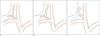
Deslaugiers et al. (28) reported that the EJV usually enters into the jugulo-subclavian venous confluence (type A, 60%), into the SCV at a distance from its junction with the IJV (type B, 36%) or into the trunk of the IJV (type C, 4%) (Fig. 1). We speculate that the course of the hemodialysis catheter using the right EJV shows a more obtuse angle than that using IJV.
We could avoid deep neck puncture by use of the EJV instead of the IJV. Although the use of real-time ultrasound guidance can reduce the risk of complications during IJV puncture, a deep puncture in the neck could lead to procedure-related complications such as arterial puncture or massive hemorrhage, especially in patients with an anatomic variation or an abnormal deep location of the IJV (29). There were no complications of massive hemorrhage or arterial puncture during EJV catheterization in this study. When bleeding occurred at the insertion site of the vein, we could easily compress the EJV because of its superficial location.
Usually, the SCV approach has a higher complication rate compared to the IJV approach. Even if the EJV is connected to the SCV (type C), we speculate that the EJV approach is different from the SCV approach since the catheter does not pass through the narrow space between the clavicle and first rib, and the right EJV has a straight course to the atrium that is similar to the right IJV.
We also think that the right EJV is a superior access route compared to the left IJV. Several studies have reported that the left IJV approach had relatively higher rates of stenosis and thrombosis compared to the right IJV approach (113143031). Our thinking is that the catheter passes through the narrow space between the sternum and aortic arch, and can cause injury or may induce stenosis by close approximation of the catheter tip to the wall of the superior vena cava.
This study has limitations for the evaluation of late complications since a hemodialysis catheter was used only until arteriovenous fistula maturation. Furthermore, the catheter dwell time was relatively short in this study.
The EJV approach could preserve the IJV from catheterization, especially in patients with end stage renal disease with a limited number of usable veins for catheterization. We suggest that the dilated right EJV could be considered as the preferred primary for hemodialysis catheter placement based on its ease of access and an acceptable complication rate. However, further prospective case-control studies are needed to clarify this.
 XML Download