

 PDF
PDF ePub
ePub Citation
 Print
Print


The association between anomalous pulmonary venous drainage and acquired valvular heart disease has rarely been reported (123456). Nonetheless, awareness of this entity is important because the appropriate surgical approach can then be identified based upon the preoperative diagnosis.
The purpose of this case report is to present a case of rheumatic mitral stenosis and partial anomalous pulmonary venous connection to the left innominate vein accompanied by the presence of the levoatriocardinal vein. The diagnosis was established preoperatively by CT and MR, thereby making complete surgical correction feasible.
Case Report
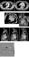
A 67-year-old woman, with a known case of mitral stenosis, underwent chest CT scanning for the preoperative evaluation of a palpable soft-tissue lesion on her chest wall. A CT scan was performed using a 16-channel multidetector CT (Sensation 16, Siemens Healthcare, Forcheim, Germany) without cardiac gating and obtained 40 sec after administration of 100 mL of a 300 mgI/mL IV contrast medium at a rate of 2.5 mL/sec. The CT parameters were as follows: 16 × 1.5 mm collimation, 100 effective milliamperes (mAs), 120 kVp, a rotation time of 0.5 seconds and pitch of 1.5. CT data were reconstructed with a 5 mm slice thickness using a soft kernel. The CT scan revealed an abnormal vascular structure in the left upper mediastinum, which connected the left innominate vein and the left atrium (levoatriocardinal vein). At the level of the aortic arch, the abnormal vessel was located on the left side of the aortic arch (Fig. 1A) and was then directed downward lateral to the aortic arch and entered through the left atrium. Before entering into the left atrium, the left superior pulmonary vein conjoined this anomalous vein, which constitutes a partial anomalous pulmonary venous return. At the level of the main pulmonary artery, this anomalous vein was located in between the left pulmonary artery and the left main bronchus (Fig. 1B). Because of its location dorsal to the pulmonary artery, this abnormal vessel could be diagnosed as a levoatriocardinal vein. On echocardiography, the patient showed tight mitral stenosis (mitral valvular area 1.0 cm2 on two-dimensional planimetry with a mean pressure gradient of 12 mmHg) with moderate to severe mitral regurgitation (jet area: 9-12 cm2).
For further evaluation of the shunt via partial anomalous pulmonary venous return, MRI was performed. Cardiac MR was performed using the Gyroscan Intera 1.5 T scanner (Philips Medical Systems, Best, The Netherlands). Balanced turbo field echo (TFE) images of the thorax were obtained from the aortic arch to the cardiac apex for anatomic delineation. The scan parameters of cine images consisted of slice thickness 8 mm, no gap, echo time/repetition time (TE/TR) of 1.7/3.4 ms, flip angle of 50 degrees, 169 phase encoding steps, and a scan field of view (FOV) measuring 320 × 320 mm. Cine images were performed for the evaluation of valvular motion. The scan parameters of cine images consisted of a slice thickness measuring 8 mm, no gap, TE/TR 1.7/3.4 ms, a flip angle of 50 degrees, 169 phase encoding steps, a scan FOV measuring 320 × 320 mm, and retrospective gating with 20 calculated phases. The flow of the superior vena cava (SVC) and the levoatriocardinal vein were obtained using velocity-encoded cine (VENC) MR imaging. The scan parameters of cine images consisted of a slice thickness measuring 8 mm, no gap, TE/TR 2.8/4.7 ms, a flip angle of 15 degrees, 128 phase encoding steps, a scan FOV measuring 320 × 320 mm, and retrospective gating.
Four-chamber cine MR images showed thickening and doming of the mitral valve leaflets, and also demonstrated a dark jet flow through the stenotic mitral valve area, which is consistent with tight mitral stenosis (Fig. 1C). Coronal MR images grossly determined the course of the levoatriocardinal vein connecting the left innominate vein and the left atrium (Fig. 1D). Sagittal MR images demonstrated that the left superior pulmonary vein connected to the levoatriocardinal vein as a form of partial anomalous pulmonary venous return (Figs. 1E-G). Hemodynamically, as the left atrial pressure increased, owing to mitral stenosis, a large amount of the left-to-right shunt was produced from both the left atrium and the left superior pulmonary vein to the left innominate vein through the levoatriocardinal vein. Assessment of the flow dynamics, including quantification of the shunt, was performed by VENC MR imaging. On phase contrast imaging, the flow direction of the SVC and the levoatriocardinal vein were opposing (Fig. 1H). On flow measurement, the ascending aorta and both central pulmonary arteries showed a flow of 40 ml/min and 100 mL/min, respectively. According to the flow measurement, the calculated left-to-right shunt was approximately 60% and the ratio of pulmonary to systemic blood flow (Qp/Qs) was approximately 2.51.
The preoperative diagnosis was mitral stenosis and partial anomalous pulmonary venous connection to the left innominate vein accompa nied with the presence of the levoatriocardinal vein. The patient underwent an open mitral commissurotomy and ligation of the levoatriocardinal vein and was ultimately discharged without any adverse events.
Discussion
The association of anomalous pulmonary venous drainage with mitral valve disease has rarely been recognized. Several cases of mitral stenosis with partial pulmonary venous return have been described in previous reports (123456). However, many of these cases showed the partial anomalous pulmonary drainage into a systemic vein, but not into an anomalous vein connecting the left atrium and the systemic vein simultaneously (456). In the present case, the left superior pulmonary vein drained into the anomalous vasculature, connecting the left atrium and the left innominate vein.
The levoatriocardinal vein, first described by Edward et al, is very uncommon and its presence is pathologic (78). It is probably derived from the persistence of anatomic channels connecting the pulmonary capillary plexus to the cardinal veins in the embryonic foregut. It is a pulmonary-systemic connection that provides an alternative pathway for pulmonary venous drainage in the presence of a severe left-sided obstructive lesion such as mitral atresia, because this unusual vein connects the left atrium or pulmonary vein to the left innominate vein.
Another anomalous vein with many similarities to the levoatriocardinal vein is a persistent left superior vena cava (PLSVC). A PLSVC is usually connected to the coronary sinus, and rarely to the left atrium or pulmonary vein (3). The levoatriocardinal vein differs from a PLSVC draining into the left atrium in that the levoatriocardinal vein ascends dorsal to the left pulmonary artery whereas the PLSVC ascends ventral to it. Moreover, the levoatriocardinal vein may be compressed between the left pulmonary artery and the left bronchus (8).
Partial anomalous pulmonary venous return (PAPVR) is most commonly seen in patients with an atrial septal defect. When the atrial septum is intact, mitral stenosis of congenital or acquired origin is the next most frequent combination (14). The association of PAPVR and rheumatic mitral stenosis appears to be no more than coincidental (2). Previous studies reported that the amount of left-to-right shunt was insignificant in patients with PAPVR, mitral stenosis, and intact interatrial septum (9). However, in our patient, the left-to-right shunt increased to as much as 60% of the pulmonary blood flow, which is the reason why the surgical ligation of the levoatriocardinal vein was performed.
This case is the first in which the preoperative diagnosis was established by MR in a patient with partial anomalous pulmonary venous return via a levoatriocardinal vein in association with rheumatic mitral stenosis. Preoperative cardiac cine MR with VENC demonstrated the anatomic configuration of the anomalous vein and allowed a quantitative assessment of the flow dynamics of the left-to-right shunt via the anomalous vein. Velocity-encoded cine MR imaging is recognized as a valuable technique for the quantitative assessment of flow dynamics in congenital heart diseases. We used this technique to measure the left-to-right shunt flow in this patient as it provided necessary hemodynamic information before surgery.
In summary, we report a rare case of partial anomalous pulmonary venous return via a levoatriocardinal vein in association with rheumatic mitral stenosis. We used MR for preoperative imaging to delineate the complicated anatomic structure and to determine the proper surgical treatment.
 XML Download