

 PDF
PDF ePub
ePub Citation
 Print
Print


Abstract
Purpose
We wanted to compare the CT findings and clinical features of parotitis and submandibular sialadenitis in children and adults and to evaluate the statistical significance of these in different age groups and the usefulness of a CT scan.
Materials and Methods
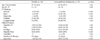
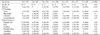
Ninety-seven adults and 36 pediatric patients with sialadenitis were included in this retrospective study. Regardless of the site of involvement, we evaluated the CT findings and clinical manifestations between the pediatric and adult groups, and between the pediatric and adult parotitis and submandibular sialadenitis groups. At last, all the patients were classified into seven age groups.
Results
Abscess formations were more prominent in the parotitis groups, and sialiths were more common in the submandibular sialadenitis group with the lowest incidence in the young children group (≤ 10 years). Cellulitis seen on a CT scan showed a higher incidence in the adult parotitis group, and this finding was closely connected with pain. A number of patients showed cervical lymphadenitis on a CT scan and this coincided with lymph node palpation. Tonsillitis associated sialadenitis was common in the pediatric group. The therapeutic durations were longer in the pediatric parotitis patient group and the adult submandibular sialadenitis group.
Figures and Tables
Fig. 1
Bilateral submandibular sialadenitis and cellulitis in a 18-year-old girl with diffuse soft tissue swellings. Postcontrast CT scan shows homogeneous enhancement and enlargement in both submandibular glands with fat infiltration (arrow) in both neck portions.
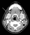
Fig. 2
Right submandibular abscess and multiple lymph nodes in a 64-year-old man.
Postcontrast CT scan shows an abscess pocket (long arrow) in right submandiublar gland with multiple cervical lymph node enlargements (short arrows).
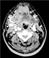
Fig. 3
Right submandibular sialadenitis and sialoliths in a 67-year-old man with postprandial pain in right neck.
A. Precontrast CT scan shows both submandublar stones (black arrows).
B. Postcontrast CT scan shows small abscess pockets (white arrows) in right submandibular gland.
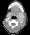
Fig. 4
Acute parotitis with tonsillitis in a 17-year-old girl with palpable mass in left infraauricular neck. Postcontrast CT scan shows left palatine tonsil (black arrow) with diffuse enlargement and enhancement in left parotid gland (white arrow).
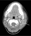
Fig. 5
Parotid abscess in a 28-year-old man with palpable mass in right infraauricular neck.
Postcontrast CT scan shows solitary abscess (arrow) in right parotid gland.
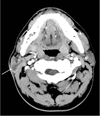
Fig. 6
Left submandibular sialadenitis in a 17-year-old girl with diffuse soft tissue painful swelling. Left submandibular gland shows diffuse enlargement and homogenous enhancement on postcontrast CT scan with cervical lymphadenitis and cellulitis.
References
1. Sumi M, Izumi M, Yonetsu K, Nakamura T. The MR imaging assessment of submandibular gland sialadenitis secondary to sialolithiasis: correlation with CT and histopathologic findings. AJNR Am J Neuroradiol. 1999; 20:1737–1743.
2. Even TE, Niv A, Kraus M, Nash M. Candida parotitis with abscess formation. Acta Otolaryngol. 2006; 126:334–336.
3. Kaneta T, Minami M, Ozawa K, Akimoto Y, Kawana T, Yamamoto H, et al. MR of the submandibular gland: normal and pathologic states. AJNR Am J Neuroradiol. 1996; 17:1575–1581.
4. Chung MK, Jeong HS, Ko MH, Cho HJ, Ryu NG, Cho DY, et al. Pediatric sialolithiasis: what is different from adult sialolithiasis? Int J Pediatr Otorhinolaryngol. 2007; 71:787–791.
5. Waseem Z, Forte V. An unusual case of bilateral submandibular sialolithiasis in a young female patient. Int J Pediatr Otorhinolaryngol. 2006; 69:691–694.
6. Bova R, Walker P. Neonatal submandibular sialadenitis progressing to submandibular gland abscess. Int J Pediatr Otorhinolaryngol. 2000; 53:73–75.
7. Bryan RN, Miller RH, Ferreyro RI, Sessions RB. Computed tomography of the major salivary glands. AJR Am J Roentgenol. 1982; 139:547–554.
8. Mandel L, Bijoor R. Imaging (computed tomography, magnetic resonance imaging, ultrasound, sialography) in a case of recurrent parotitis in children. J Oral Maxillofac Surg. 2006; 64:984–988.
9. Mandel L, Hatzis G. The role of computerized tomography in the diagnosis and therapy of parotid stones: a case report. J Am Dent Assoc. 2000; 131:479–482.
10. Laskawi R, Schaffranietz F, Arglebe C, Ellies M. Inflammatory diseases of the salivary glands in infants and adolescents. Int J Pediatr Otorhinolaryngol. 2006; 70:129–136.
11. Becker M, Marchal F, Becker CD, Dulguerov P, Georgakopoulos G, Lehmann W, et al. Sialolithiasis and salivary ductal stenosis: diagnostic accuracy of MR sialography with a three-dimensional extended-Phase conjugate-symmetry rapid spin-echo sequence. Radiology. 2000; 217:347–358.
12. Stong BC, Sipp JA, Sobol SE. Pediatric parotitis: a 5-year review at a tertiary care pediatric institution. Int J Pediatr Otorhinolaryngol. 2006; 70:541–544.
13. Sitheeque M, Sivachandran Y, Varathan V, Ariyawardana A, Ranasinghe A. Juvenile recurrent parotitis: clinical, sialographic and ultrasonographic features. Int J Paediatr Dent. 2007; 17:98–104.
14. Miziara ID, Campelo VE. Infantile recurrent parotitis: follow up study of five cases and literature review. Braz J Otorhinolaryngol. 2005; 71:570–575.
15. Saarinen RT, Kolho KL, Pitkaranta A. Cases presenting as parotid abscesses in children. Int J Pediatr Otorhinolaryngol. 2007; 71:897–901.
16. Faure F, Querin S, Dulguerov P, Froehlich P, Disant F, Marchal F. Pediatric salivary gland obstructive swelling: sialendoscopic approach. Laryngoscope. 2007; 117:1364–1367.
17. Nahlieli O, Eliav E, Hasson O, Zagury A, Baruchin AM. Pediatric sialolithiasis. Oral Surg Oral Med Oral Pathol Oral Radiol Endod. 2000; 90:709–712.
 XML Download