

 PDF
PDF ePub
ePub Citation
 Print
Print


Spontaneous rupture of the liver is rare, but it can be the most serious and potentially life-threatening complication associated with preeclampsia or eclampsia. The patient's survival generally depends on the early recognition of the characteristic signs and symptoms and prompt surgical intervention. Even with surgical intervention, the maternal mortality had been reported to be as high as 59-86% (12). We describe here a case of successful treatment of spontaneous hepatic rupture due to preeclampsia of pregnancy by performing transcatheter hepatic arterial embolization, and we also review the relevant literature.
Case Report
A 41-year-old woman (gravida 3, para 2) at 34 weeks gestation presented with epigastric pain. Her blood pressure was noted to be 178/100 mmHg with 2+ proteinuria, and we observed signs of fetal distress. So, she underwent an emergency cesarean section and she delivered a live healthy female infant.
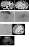
Five hours after delivery, she developed severe epigastric pain without any vaginal bleeding. Her hematocrit fell from 48% to 20%. Her blood pressure fell from 178/100 mmHg to 80/40 mmHg. Massive transfusions were begun and an emergency CT scan was performed. An emergency CT scan showed a large, poorly circumscribed, low density area in the both lobes of the liver, which contained irregular frond-like branches of normal hepatic density. A moderate amount of adjacent subcapsular fluid was present, as well as a moderate amount of free intraperitoneal fluid (Figs. 1A, B). Massive transfusions were begun and an emergency hepatic angiogram was obtained.
The emergency hepatic angiogram demonstrated displacement of hepatic vessels due to a large subcapsular hematoma and multiple small punctate areas of extravasation in both lobes of the liver (Figs. 1C, D). We then performed angiographic embolization of both hepatic arteries with using a 2.7-French microcatheter (Progreat, Terumo, Tokyo, Japan). Gelfoam pledge were injected into both the hepatic arteries until the arterial blood flow to the liver was completely blocked (Fig. 1E).
After embolization of the hepatic arteries, the patient's vital signs stabilized and her hematocrit remained stable.
A computed tomographic (CT) scan 10 days after embolization showed a large amount of subcapsular hematoma of the liver and some intraperitoneal free fluid (Fig. 1F). Eleven days after embolization, she had a low-grade fever and abdominal fullness, so insertion of pigtail drainage catheter was performed and the extensive subcapular hematoma from the liver was drained. The patient recovered gradually from her abdominal fullness and her persistent low grade fever. Her recovery from this procedure was uneventful, and she was discharged 4 weeks later in good condition. The follow-up ultrasonogram that was done 8 weeks after embolization in the outpatient department showed the barely visible subcapsular hematoma of the liver and the intraabdominal hematoma (Fig. 1G).
Discussion
Spontaneous rupture of liver is extremely rare, and it can occur in association with hepatocelluar carcinoma, hepatic hemangioma, hepatic abscess or rarely pregnancy (1). When it is associated with pregnancy, it has especially occurred combined with eclampsia or preeclampsia.
Rupture of liver associated with eclampsia or preeclampsia generally occurs in a multiparous patient with an average age of the late 20s or early 30s. The vast majority of cases have occurred in the third or late second trimester of pregnancy, although approximately 15% of the reported cases have occurred in the puerperium, and usually within 24 hours of delivery, as in the present case (2). Generally, there are signs of preeclampsia present when the patient experiences a sudden onset of epigastric pain or moderate to severe right upper quadrant abdominal pain and eventually hypovolemic shock (3).
The pathogenesis of spontaneous rupture of liver associated with eclampsia or preeclampsia is unclear. Eclamptic patients are generally prone toward spontaneous hemorrhage and they frequently show fibrin deposition in the hepatic sinusoids and arterioles. Microangiopathic changes that are the consequence of fibrin deposition result in the occlusion of the hepatic sinusoids and necrosis of the small arterioles within the liver. As a result, infarction with vascular disruption may occur, leading to intrahepatic hemorrhage, rupture of the parenchyma, production of a subcapsular hematoma and ultimately rupture of Glisson's capsule with intraperitoneal hemorrhage (4). Some of the hepatic lesions in eclampsia have been attributed to disseminated intravascular coagulation (DIC) (5). McKay describes that eclampsia is actually a form of disseminated intravascular coagulation (6), yet others feel that DIC is not the primary event. In our case and in other reported cases, multiple small pseudoaneurysms were present in the area of hemorrhage, which is consistent with the suggestion that a toxic vasculopathy is the cause of this rare complication of pregnancy (7).
Survival appears to be closely related to making a rapid diagnosis and prompt surgical intervention. Of the currently available imaging techniques such as high resolution ultrasound examination and computed tomography examination for confirming the diagnosis of hepatic rupture, computed tomography is by far the most useful if the patent is relatively stable.
Conservative treatment of the contained intraparenchymal or subcapsular hemorrhage without free intraperitoneal rupture has been performed without subsequent death, but surgical intervention is thought to improve survival. A number of methods, including packing, hepatic resection, hepatic artery occlusion by ligation, hepatic lobectomy and liver transplantation have been employed to achieve hemostasis. But there is a difficulty to achieve liver hemostasis surgically because of the presence of multiple areas of infarction and hematoma, and especially in the presence of coagulopathy.
Transcatheter embolization of the hepatic artery for controlling hemorrahge and for treating hepatic neoplasms has been well described (89). Several recently reported cases of successful management with hepatic artery embolization of hepatic rupture associated with pregnancy have been described (31011). We performed transcatheter embolization of the hepatic artery as a mean of obtaining hemostasis. We used 1 mm cubes of Gelfoam sponge for our patient. Gelfoam seems to be the ideal embolic material as it is a gelatin sponge that is resorbed over several weeks, and so it usually allows partial recanalization of the occluded vessels. We performed embolization with the catheter tip in the proximal right hepatic artery and left hepatic artery. Because there was diffuse bleeding in both lobes, selective catheterization was not attempted. Embolization of the hepatic artery is a relatively safe procedure because the liver has a dual blood supply from the portal vein and hepatic artery.
In conclusion, transcatheter hepatic artery embolization is a safe, effective alternative to surgical treatment and it can play a major role in controlling hepatic rupture associated with preeclampsia.
 XML Download