

 PDF
PDF ePub
ePub Citation
 Print
Print


Desmoplastic fibroblastoma (collagenous fibroma) is a fibrous, soft-tissue tumor first described by Evans (1) in 1995. This tumor is clinically and morphologically distinct and was deemed completely benign in a few reported series (123). There have also been published case reports on the MR features of desmoplastic fibroblastoma (45678). We herein present a case of desmoplastic fibroblastoma with special focus on the MR findings, because they are distinct from that previously published.
Case Report
A 33-year-old female presented with an eight-month history of a palpable mass in her right arm without associated neurologic symptoms. Physical examination revealed a hard, well-circumscribed and movable mass measuring 4×4×2 cm. Plain radiography of the mass demonstrated no calcification (not shown).
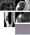
Gray-scale imaging on ultrasonography showed a well-defined, hypoechoic mass in the superficial portion of the muscle layer of the patient's right arm. Color Doppler imaging revealed vascular signals in the peripheral and central portions of the mass (Fig. 1A). MR imaging was then performed. The mass is located in the subcutaneous layer and is attached to the right biceps brachii and brachialis muscles. Axial fast spin echo (FSE) T1-weighted images of the mass revealed slightly higher signal intensity, compared to that of the adjacent muscles (Fig. 1B). Coronal FSE T2-weighted images of the mass revealed slightly lower signal intensity, with higher signal intensity in the upper area of the mass (Fig. 1C). Axial, post-contrast T1-weighted images showed marked enhancement (Fig. 1D). The mass showed mild indentation to the adjacent muscles with no evidence of invasion.
The mass was easily and completely removed by excisional biopsy. There was no evidence of invasion to the muscles of her right arm, and there were no tumor cells in the resected margin of the frozen section.
Macroscopically, the tumor was firm without cystic or hemorrhagic changes. Microscopically, the tumor was composed of spindle to stellate fibroblastic cells in a hypovascular collagenous matrix (Fig. 1E). The immunohistochemical study was negative on S-100 protein. The final diagnosis was desmoplastic fibroblastoma.
The patient experienced no signs of recurrence and remained healthy 16 months after surgery.
Discussion
Desmoplastic fibroblastoma (collagenous fibroma) is a benign fibrous tumor most often seen in patients during the fifth and sixth decades of life, with a male to female ratio of 5:1. Most tumors occur in the extremities, shoulder girdle, posterior neck, upper back and abdominal wall. The size of reported desmoplastic fibroblastomas ranges from 1 to 20 cm in maximal diameter. The lesion is typically a well-circumscribed, firm mass that involves the subcutaneous or deep soft tissue (123).
Histologically, the tumor cells are relatively bland stellate- and spindle-shaped fibroblastic cells, separated by a densely fibrous to fibromyxoid matrix. The cellularity ranges from low to very low. The mitotic figures are very low or completely absent. Tumor necrosis is not seen (123).
There have been a few case reports of desmoplastic fibroblastoma with MR features (45678). Three cases of desmoplastic fibroblastoma in the upper extremity, and one case in a hip joint showing diffusely low signal intensity of the mass on T2-weighted images (457) have been reported. One reported case of desmoplastic fibroblastoma in the peritoneal cavity showed marked heterogeneous signal intensity on T2-weighted and post-contrast T1-weighted images (6). These findings are slightly different from that seen in our case, which had heterogeneously high and low signal intensity on T2-weighted images, and well-enhanced post-contrast T1-weighted images. According to a recent report of desmoplastic fibroblastoma with erosion of the right L5 pedicle, MR images showed heterogeneous intermediate signal intensity on T2-weighted image with scattered areas of low signal intensity, and minimally heterogeneous enhancement (8). However, our case showed higher heterogeneous signal intensity on T2-weighted images and more prominent enhancement.
Most soft-tissue masses have high signal intensity on T2-weighted images. In the absence of calcification, abundant collagen and marked hypocellularity in a soft-tissue tumor resulted in reduced signal on T2-weighted pulse sequence (9). The area showing high signal intensity on T2-weighted images corresponded to the hypercellular area within the lesion, consisted of a tumor with loose collagen fibers (6). The high signal intensity on T2-weighted images seen in our case is probably due to high cellularity and small amount of loose collagen fibers in the mass.
The radiologic differential diagnosis included other soft-tissue neoplasms, such as desmoid tumor, neurogenic tumor and soft-tissue sarcoma. Among these differential diagnosis, desmoid tumor is the most important consideration for clinicians. Histologically, desmoid tumor has a greater infiltrative potential and is usually more cellular than desmoplastic fibroblastoma. On MR imaging, desmoid tumor may also have larger areas showing very high signal intensity on T2-weighted images, thus indicating higher cellularity than desmoplastic fibroblastoma (10). If high signal intensity on T2-weighted images is prominent on MR imaging, as was in our case, differentiating desmoplastic fibroblastoma from desmoid tumor becomes very difficult.
Although desmoplastic fibroblastoma is usually revealed by low signal intensity on T2-weighted MR images, it may appear as a well-defined soft-tissue mass with heterogeneously high and low signal intensity on T2-weighted images, and as intense enhancement on MR imaging
 XML Download