

 PDF
PDF ePub
ePub Citation
 Print
Print


Abstract
Peripheral primitive neuroectodermal tumors (peripheral PNETs) are very rare and highly aggressive soft-tissue malignancies originating from the neural crest. To the best of our knowledge, only a few cases of peripheral PNETs of the stomach have been reported in the literature. We report a case of large peripheral primitive neuroectodermal tumor of the stomach with MDCT findings in a 22-year-old man presenting epigastric pain and vomiting.
Figures and Tables
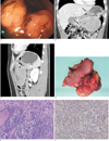
Fig. 1
Peripheral primitive neuroectodermal tumor of the stomach in a 22-year-old man.
A. Photograph from conventional gastroscopy shows a large submucosal mass arising from the gastric body and antrum.
B, C. Coronal and sagittal MPR images of contrast-enhanced MDCT shows a large lobulated solid mass arising from the gastric body and antrum with endogastric and exogastric growth and relatively smooth outer border. Note two regional lymph nodes metastases (arrows) in the perigastric space.
D. Photograph of gross specimen shows a large lobulated mass (12×9 cm in size) with invasion to the whole layer of gastric wall and extensive perigastric fat tissue.
E. Photomicrograph shows small round cells with invasion to the submucosal layer of gastric wall and extensive necrosis. Note distinctive lobular architecture with fibrous septa (H & E staining, ×40).
F. Photomicrograph of an immunohistochemistry for detecting MIC2 protein (CD99) shows intense tumor cells membranous immunoreactivity (×400).
References
1. Kimber C, Michalski A, Spitz L, Pierro A. Primitive neuroectodermal tumours: anatomic location, extent of surgery, and outcome. J Pediatr Surg. 1998; 33:39–41.
2. Soulard R, Claude V, Camparo P, Dufau JP, Saint-Blancard P, Gros P. Primitive neuroectodermal tumor of the stomach. Arch Pathol Lab Med. 2005; 129:107–110.
3. Katz RL, Quezado M, Senderowicz AM, Villalba L, Laskin WB, Tsokos M. An intra-abdominal small round cell neoplasm with features of primitive neuroectodermal and desmoplastic round cell tumor and a EWS/FLI-1 fusion transcript. Hum Pathol. 1997; 28:502–509.
4. Inaba H, Ohta S, Nishimura T, Takamochi K, Ishida I, Etoh T, et al. An operative case of primitive neuroectodermal tumor in the posterior mediastinum. Kyobu Geka. 1998; 51:250–253.
5. Amin HM, Candel AG, Husain AN. Pathologic quiz case. A 22-year-old man with an abdominal mass. Arch Pathol Lab Med. 1999; 123:541–543.
6. Gasecki D, Izycka-Swieszewska E, Szymkiewicz-Rogowska A, Kopczynski S, Mechlinska-Baczkowska J. Primitive neuroectodermal tumor of rare localization in two members of one family. Neurol Neurochir Pol. 1999; 33:1415–1423.
7. Ibarburen C, Haberman JJ, Zerhouni EA. Peripheral primitive neuroectodermal tumors. CT and MRI evaluation. Eur J Radiol. 1996; 21:225–232.
8. Kim MS, Kim B, Park CS, Song SY, Lee EJ, Park NH, et al. Radiologic findings of peripheral primitive neuroectodermal tumor arising in the retroperitoneum. AJR Am J Roentgenol. 2006; 186:1125–1132.
9. Peter M, Gilbert E, Delattre O. A multiplex real-time PCR assay for the detection of gene fusions observed in solid tumors. Lab Invest. 2001; 81:905–912.
10. Ushigome S, Machinami R, Sorensen PH. In Ewing sarcoma/primitive neuroectodermal tumour (PNET). In : Fletcher CDM, Unnik K, Mertens F, editors. World Health Organization Classification of Tumours Pathology and Genetics of Tumours of Soft Tissue and Bone. Lyon, France: IARC Press;2002. p. 297–300.
 XML Download