

 PDF
PDF ePub
ePub Citation
 Print
Print


Melorheostosis is a rare nonhereditary sclerosing mesenchymal dysplasia of bone that is diagnosed by a characteristic linear cortical hyperostosis with a flowing candle wax appearance (123). It commonly affects long bones, usually in the lower limb and on one side of the body. The involvement of flat bones is rare (12). In addition, bilateral involvement is extremely rare, with only one case of long tubular bones with a symmetric distribution reported to date. We present the first case of melorheostosis with multiple flat bone involvement and symmetric distribution of the lesions.
Case Report
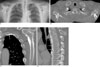
A 55-year-old man was diagnosed with sclerotic ribs and scapulas on a chest radiograph (Fig. 1A) as part of a routine check-up. Neither the patient nor his family members had a history of significant disease. The patient's blood chemistry and physical examination revealed no significant abnormalities. A chest CT showed uneven cortical thickening in multiple ribs, both scapulas, and thoracic vertebral bodies. The cortical thickening of the ribs and scapulas was bilateral and symmetrical in distribution (Fig. 1B). Our differential diagnosis included the polyostotic form of melorheostosis, bilateral osteoma, and osteoblastic metastasis. For further evaluation, using the acquired volume data, we obtained multiplanar reconstruction (MPR) images, which revealed that cortical thickening was seen along the lower aspects of the ribs, infraspinatus fossas, the spines of both scapulas, and the anterior column of the thoracic vertebral bodies (Figs. 1C, D).
Discussion
Melorheostosis is a rare, osteosclerotic dysplasia with an unknown etiology and first described in 1922 by Leri and Joanny (12). The diagnosis is based on the clinical and radiological findings. The radiological appearance of melorheostosis is a characteristic wavy cortical hyperostosis along the surfaces of the long bone with vertical extension, and resembles melted wax dripping down one side of a candle; the so called flowing candle wax appearance (24). Melorheostosis frequently involves the long tubular bones. By far, the most common sites affected include the long bones of the lower limbs (4). This disorder tends to be segmental and unilateral, and based on the extension of the bone involvement of the lesion, it may be monostotic, monomelic, or polyostotic (4). A bone scintigraphy is invariably positive in patients with melorheostosis, revealing a moderate increase in uptake of tracer, predominantly localized to the cortex (34). Computed tomography (CT) scanning more effectively reveals a clear demarcation from normal bone than standard radiographs (4). Abnormalities of the soft tissues overlying the bone and joint lesions are common, and in many cases, associated scleroderma have been reported (34).
Most of the reported cases showed distribution of one long bone or one limb with a scleroderma distribution (5). This characteristic distribution pattern and radiological features differentiates melorheostosis from other osteosclerotic dysplasias. Isolated flat bone involvement is uncommon (3), and multiple flat bone involvement without long bone involvement, as seen in our patient, is exceedingly rare. Moreover, symmetric involvement of multiple flat bones has not been previously reported.
Melorheostosis is a rare dysplasia with characteristic cortical thickening, commonly involving the long bones of one limb. Here we report the first case of melorheostosis with the characteristic CT findings and an unusual distribution pattern with multiple flat bones that were symmetrically involved without long bone involvement.
 XML Download