

 PDF
PDF ePub
ePub Citation
 Print
Print


Abstract
Purpose
To predict the histopathologic grading of liposarcomas with the modified classification of MRI findings.
Materials and Methods
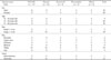
The 47 MRI studies of pathologically confirmed liposarcomas were retrospectively analyzed, and found to have well-differentiated (n=17) and myxoid (n=16) types, representing the 'low grade' sarcomas, as well as the round cell (n=9) and pleomorphic (n=5) types, which represent the 'high grade' sarcomas. The MRI findings of liposarcomas were classified into four groups as the one with fat signal above 80%, 20-80%, below 20%, and no fat signal on T1-weighted images (WIs), into three groups as the one with high signals with reticular or irregular lines, homogeneous above 50% of the area, and heterogeneous above 50% on the T2WIs, and into three groups as the one enhanced with reticular or irregular lines and enhancement areas above 50% and below 50% on the Gd-DTPA T1WIs.
Figures and Tables
Fig. 1
83-year-old woman with well-differentiated liposarcoma in the vastus muscles of the right thigh.
A. Coronal T1-weighted spin-echo MR image (WI) reveals near-total high signal intensity tumor as fat mass above 80% (large arrows) ('A1').
B. Coronal T2-WI with fat saturation (FS) reveals central reticular high signal lesions (small arrows) ('B1').
C. Coronal contrast-enhanced T1-WI with FS shows reticular and thick high signal lesions (small arrows) ('C1').
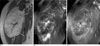
Fig. 2
40-year-old woman with multiple myxoid liposarcomas in the semitendinous muscle of the right thigh.
A. Coronal T1-weighted spin-echo MR image (WI) reveals peripheral focal lacy high signal area as fat below 20% (arrow) ('A3').
B. Coronal T2-WI with fat saturation (FS) reveals homogeneous high signal area above 50% (arrows) ('B2').
C. Coronal contrast-enhanced T1-WI with FS shows whirpool like high signal areas (arrows) ('C2a').
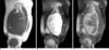
Fig. 3
44-year-old man with round cell liposarcoma in the soleus muscle at the right calf.
A. Axial T1-weighted spin-echo MR image (WI) reveals large iso-signal intensity mass with hemorrhagic foci (arrows) ('A4').
B. Axial T2-WI reveals relatively homogeneous high signal above 50% of the tumor ('B2').
C. Axial contrast-enhanced T1-WI with fat saturation shows enhancement area below 50% of the tumor (arrows) ('C3').
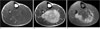
Fig. 4
59-year-old man with pleomorphic liposarcoma in the gastrocnemius muscle of the left calf.
A. Axial T1-weighted spin-echo MR image (WI) reveals large iso-signal intensity mass with hemorrhagic foci (arrows) instead of fat focus ('A4').
B. Axial T2-WI reveals relatively homogeneous high signal area above 50% ('B2').
C. Axial contrast-enhanced T1-WI with fat saturation shows peripheral enhancement with central avascular area (arrows) ('C3a').


References
1. Reszel PA, Soule EH, Coventry MB. Liposarcoma of the extremities and limb girdles: a study of 222 cases. J Bone Joint Surg Am. 1966; 48:229–244.
2. Kransdorf MJ. Malignant soft-tissue tumors in a large referral population: distribution of diagnoses by age, sex, and location. AJR Am J Roentgenol. 1995; 164:129–134.
3. Jelinek JS, Kransdorf MJ, Shmookler BM, Aboulafia AJ, Malawer MM. Liposarcoma of the extremities: MR and CT findings in the histologic subtypes. Radiology. 1993; 186:455–459.
4. Kim JI, Choi KU, Lee IS, Moon TY, Lee CH, Kim HW, et al. Gene expression in mixed type liposarcoma. Pathology. 2006; 38:114–119.
5. Fletcher CD, Unni KK, Mertens F. Pathology and genetics of tumours of soft tissue and bone. Lyon, France: IARC;2002.
6. Tateishi U, Hasegawa T, Beppu Y, Kawai A, Satake M, Moriyama N. Prognostic significance of MRI findings in patients with myxoid-round cell liposarcoma. AJR Am J Roentgenol. 2004; 182:725–731.
7. Arkun R, Memis A, Akalin T, Ustun EE, Sabah D, Kandiloglu G. Liposarcoma of soft tissue: MRI findings with pathologic correlation. Skeletal Radiol. 1997; 26:167–172.
8. Kransdorf MJ, Moser RP, Meis JM, Meyer CA. Fat containing soft tissue masses of the extremities. Radiographics. 1991; 11:81–106.
9. Wu JS, Hochman MG. Soft-tissue tumors and tumorlike lesions: a systematic imaging approach. Radiology. 2009; 253:297–316.
10. Orson GG, Sim FH, Reiman HM, Taylor WF. Liposarcoma of the musculoskeletal system. Cancer. 1987; 60:1362–1270.
11. Kransdorf MJ, Bancroft LW, Peterson JJ, Murphey MD, Foster WC, Temple HT. Imaging of fatty tumors: distinction of lipoma and well-differentiated liposarcoma. Radiology. 2002; 224:99–104.
12. Sung MS, Kang HS, Suh JS, Lee JH, Park JM, Kim JY, et al. Myxoid liposarcoma: appearance at MR imaging with histologic correlation. Radiographics. 2000; 20:1007–1019.
13. Enzinger FM, Weiss SW. Soft tissue tumors. 2nd ed. St Louis: Mosby;1988. p. 346–382.
14. Song T, Shen J, Liang BL, Mai WW, Li Y, Guo HC. Retroperitoneal liposarcoma: MR characteristics and pathological correlative analysis. Abdom Imaging. 2007; 32:668–674.
15. Baur A, Bartl R, Pellengahr C, Baltin V, Peiser M. Neovascularization of bone marrow in patients with diffuse multiple myeloma. Cancer. 2004; 101:2599–2604.
16. Moon TY, Lee IS, Lee G, Kim JI, Choi KU, Kim WT. MR Histoanatomical Distribution of 290 Soft-tissue Tumors. J Korean Radiol Soc. 2008; 59:417–427.
 XML Download