

 PDF
PDF ePub
ePub Citation
 Print
Print


Angiomyolipoma (AML) is a mesenchymal neoplasm composed of variable proportions of dysmorphic blood vessels, smooth muscle, and adipose tissue. AMLs are typically solid lesions both radiologically and pathologically (1). Recently, AMLs, including the gross or microscopic cystic component, have been reported in the pathologic literature and discovered as AML embedding epithelial elements in the form of cysts and termed AML with epithelial cysts (AMLEC) or cystic AML (12). There has been only one previous report about the radiologic findings of AMLEC in 2010. They described the AMLEC as a soild mass containing a tiny cystic focus (3). In this report, we present a case of AMLEC in a 67-year-old man presenting as a multilocular cystic mass without a visible solid portion and located in the kidney.
Case Report
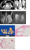
A 67-year-old man presented with a right renal mass, which was incidentally detected during CT scan as part of a staging workup for colon cancer. The man had no urinary symptom such as flank pain or hematuria. An enhanced abdominal CT scan revealed a 3.5 cm-sized exophytic, multilocular cystic mass in the lower pole of the right kidney along with an enhancing thin wall and septa (Figs. 1A, B). Moreover, there was no fat-density lesion or enhancing solid component in the mass. These findings are indicative of a Bosniak III lesion. An MR scan identified homogenous high signal intensity fluid in the locules of the multi-septated mass on a T2-weighted image, as well as enhancement of the thin wall and septa without a solid portion on a fat-suppressed enhanced T1-weighted image (Figs. 1C, D). The radiological differential diagnoses were cystic renal cell carcinoma, multilocular cystic nephroma or mixed epithelial and stromal tumor (MEST). The patient also had sigmoid colon cancer with two metastatic nodules in segment V and VIII of the liver. As a result, an anterior resection, hepatic segmentectomy and partial nephrectomy were performed.
Macroscopically, the mass was a well-demarcated multi-cystic mass on the renal cortex (Fig. 1E). The cysts had a smooth wall which contained serous fluid. Grossly, no capsular or cortical invasion was suspected.
Microscopic examination revealed that the mass contained multiple epithelial cysts and intercystic stromas, which consisted of smooth muscle bundles and adipose tissue admixed with small or medium sized vessels. These pathologic findings of stromal component were consistent with angiomyolipoma (Figs. 1F, G). The epithelial cyst lining cells showed bland flat columnar epithelium, which was positive for pancytokeratin (not shown). Altogether, the mass was diagnosed as angiomyolipoma with an epithelial cyst.
Discussion
Classic AMLs have a triphasic appearance containing all three components. Some tumors consisting almost exclusively of a single component are called monophasic AML. Other morphologic variants such as epithelioid, oncocytic, and cystic AMLs are rarely seen (4). Because AML is a mesenchymal tumor, it is thought to be lacking an epithelial component. Although the epithelial component, which presented in the form of entrapped renal tubules have been reported in AML, presentation as a cystic mass has been recently described (1). This distinct cystic variant of AML embeds epithelial cysts lined with epithelium (4). This has been termed angiomyolipoma with epithelial cysts (AMLEC) by Fine and colleagues (5) or cystic AML by Davis and colleagues (2). This is a descriptive name that does consider the pathogenesis or relationship to other neoplasms.
The nature of the epithelium within AMLEC is unclear. Fine et al. (5) have suggested that the epithelial component of AMLEC mainly represented a dilated, entrapped, native renal collecting duct epithelium. However, Davis et al. (2) reported that it represented true epithelial differentiation by the AML.
Prevalence of AMLEC is also not well known. Although it is thought that epithelium is extremely rare in AML, several abstracts have reported this occurrence (5). Leung et al. (6) reported 23 (71.8%) cases of AML with an epithelial component as entrapped tubules in 32 cases of AML. In 6 (18.7%) cases, the tubules showed variable cystic dilatation. To the best of our knowledge, 17 cases of AMLEC have been reported in four pathologic articles over the last three years (1257). Aydin et al. (4) reports 13 cases (6.7%) in a clinicopathologic study of 194 cases of AML.
Although the clinical outcome of AMLEC is not established, it is thought to be benign. There have been no reports of local recurrence or distant metastasis (1257).
On histological examination, the tumor consists of three components. The first component consists of cystic or multicystic spaces lined by flat, cuboidal to columnar epithelia. The second component is a compact subepithelial stroma. And, the third component includes AML elements such as smooth muscle, dysmorphic vessels, or adipose tissue. Almost all reported cases of AMLEC contained minimal to no fat (1257), which indicates that AMLEC may be a cystic variant of myomatous AML (3). However, in one case, "about 20%" fat was noted (2), and our case also showed adipose tissue, microscopically. Specifically, subepithelial stroma and exterior smooth muscle in AMLs express melanocytic markers including HMB45 and melan A, which are diagnostic of AML.
In a previously reported case in a radiologic journal, AMLEC was demonstrated as a soild mass containing a tiny cyst (3). To date, pathologic reports have indicated that AMLEC has demonstrated various gross features including a cystic mass with a mural nodule, a solid mass with an internal cyst, or a septated cyst (1257). Of these, septated cystic masses were reported in two cases (27). Our patient presented with a multi-loculated cystic mass without a fat or solid component. The wall and septa of the mass was smooth and thin. In this case, the differential diagnoses usually include cystic renal cell carcinoma (RCC), multilocular cystic nephroma (MLCN), and mixed epithelial and stromal tumor (MEST). Imaging features such as enhancing solid tissues, intratumoral hemorrhage, or thick and nodular septal enhancement may favor RCC. Findings suggestive of MLCN include herniation of a portion of the mass into the renal pelvis. MEST has a female predominance and association with hormone therapy. In most cases, if there are no supportive findings, it is not possible to definitively differentiate these disease entities radiologically (8). Thus histologic confirmation is necessary to establish the differential diagnosis.
In summary, although AMLEC is uncommon and shows no specific imaging features, it needs to be considered in the differential diagnosis of a multi-locular cystic mass arising in the kidney.
 XML Download