

 PDF
PDF ePub
ePub Citation
 Print
Print


Ectopic pancreas, also known as heterotrophic, accessory, or aberrant pancreas is a pancreatic tissue without anatomic and vascular continuity within the main body of the gland, and is most commonly found in the antrum of the stomach, duodenum, or proximal jejunum (12). Ectopic pancreas in the proximal stomach has been rarely reported (3). However, the coexistence of two ectopic pancreases at the gastric cardia and antrum in a patient has not been previously reported.
Here, we present a case of the coexistence of two ectopic pancreases at the cardia and antrum of the stomach in a 60-year-old man.
Case Report
A 60-year-old man who visited a local clinic was incidentally found to have two submucosal lesions in the cardia and antrum of his stomach on gastroscopy. Consequently, the patient came to our hospital for further evaluation and found no gastrointestinal symptoms including abdominal discomfort or pain. The patient's past medical history and initial laboratory data (including a stool occult blood test) revealed no remarkable findings.
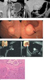
The patient underwent an abdominal computed tomography (CT) scan which was obtained before the administration of intravenous contrast material. In addition, portal venous phase images were also obtained after the administration of intravenous contrast with a 70 second delay. CT showed two gastric lesions in the gastric cardia and antrum, respectively (Figs. 1A, B). The lesion in gastric cardia appeared as a relatively well-defined heterogeneously enhancing submucosal mass with a mixed growth pattern, which was bulging into the extraluminal and endoluminal space, and had a maximum diameter of 2.2 cm (Fig. 1A). The small cystic portion in it was suspicious, but the overlying mucosa seems to be intact. The other lesion in the greater curvature side of the gastric antrum was diagnosed as an ill-defined heterogeneously enhancing mass with endoluminal growth pattern, and a maximum diameter of 2.5 cm (Fig. 1B). The internal cystic portion or overlying mucosa of it was not definite on CT. Considering the CT findings, which were double lesions with a different location and growth pattern, we could not predict the possibility of ectopic pancreas; however, we provided the possibility of a gastrointestinal stromal tumor (GIST).
Two weeks later, the patient underwent an endoscopy at our hospital. The lesion in the gastric cardia appeared as a lobular submucosal mass with intact overlying mucosa, while the other lesion in the greater curvature side of the gastric antrum appeared as a submucosal mass with a focal shallow pit at the overlying mucosa (Fig. 1C). On endoscopic ultrasonography (EUS), both lesions appeared as indistinct, heterogeneous, and intermediate hypoechoic lesions with internal anechoic cystic portion (Fig. 1D).
Although two submucosal lesions with an internal cystic portion on EUS suggested the possibility of ectopic pancreas, the possibility of GIST could not be excluded. On endoscopic biopsy, only chronic inflammatory tissue could be obtained. The patient underwent an endoscopic mucosal resection for the lesion in the greater curvature side of the gastric antrum and a surgical wedge resection of the stomach for the lesion in gastric cardia.
On gross examination, two relatively well-demarcated, firm, yellow, lobular masses (2.3 × 2.0 × 1.0 cm and 3.5 × 2.0 × 1.5 cm, respectively) were seen in the gastric wall of the antrum and cardia, respectively. The overlying gastric mucosa of the antrum showed central umbilication, but that of the cardia was intact. On microscopic examination, both masses were composed of normal pancreatic acinic and ductal tissues, consisting of ectopic pancreas (Fig. E).
Discussion
Ectopic pancreas could occur throughout the whole gastrointestinal tract. In rare cases, this anomaly is found in the gallbladder, biliary tree, liver, spleen, omentum, mesentery, appendix, mediastinum, or Meckel's diverticulum (4). About 80% of ectopic pancreases are located in the stomach, duodenum, or proximal jejunum (5). When in the stomach, ectopic pancreas typically has the appearance of a submucosal lesion in the greater curvature side of gastric antrum, 1 to 6 cm from the pylorus (4). Rarely, ectopic pancreas in the proximal stomach has been reported (36). However, to our knowledge, the coexistence of two ectopic pancreases in a patient has not been reported in the English literature. Among the various kinds of submucosal tumors of the stomach, neurofibromas, granular cell tumors, metastases, or lymphomas may appear as two or more lesions in a patient (47).
By using diagnostic imaging modalities such as barium studies or CT, it is difficult to differentiate between ectopic pancreas and other common submucosal tumors, such as GISTs or leiomyomas. Thus, ectopic pancreas is commonly mistaken for GIST (2). However, some imaging features could help to reveal ectopic pancreas. These features are typically located in the distal antrum of the stomach with occasional rudimentary ductal structures found within the lesion. Therefore, when a submucosal tumor with central umbilication located in the gastric antrum is found on a single or double contrast barium study, the likelihood of ectopic pancreas is very high (4). On CT, the finding of ill-defined submucosal lesion at its typical location, heterogeneous enhancement, and endoluminal growth pattern favor ectopic pancreas (2). Sometimes small cystic portion could be found within the ectopic pancreatic tissue due to the dilation of the anomalous duct at CT (6). At endoscopy and EUS, the discovery of a submucosal mass with central dimpling, indistinct margin, and heterogeneous echogenicity favor ectopic pancreas diagnosis (8).
Usually, asymptomatic lesions of ectopic pancreas require no further treatment after histological confirm. However, it is difficult to obtain a definitive preoperative diagnosis of ectopic pancreas and therefore a surgical resection is recommended for most patients (9). Recently, there have been several reports suggesting EMR as an attractive, less invasive option for the resection accessible lesions (10).
In summary, of the two ectopic pancreas lesions in our case, only the lesion located within the gastric antrum fits the typical CT finding of ectopic pancreas. Thus, we could not diagnose them as ectopic pancreases. Therefore, if two or more gastric submucosal lesions are coexisting in one patient, despite their different growth patterns, location, and appearance at CT, we should consider the possibility of ectopic pancreas.
 XML Download