

 PDF
PDF ePub
ePub Citation
 Print
Print


Primary synovial chondromatosis is a benign monoarticular disorder of unknown etiology characterized by non-neoplastic, proliferative and metaplastic disorder of the synovium (1).
Documented malignant transformation of primary synovial chondromatosis to chondosarcoma is probably most rare. It is not clear whether synovial chondrosarcoma arises de novo or from a malignant transformation of synovial chondromatosis (23456). Like synovial chondromatosis, synovial chondrosarcomas are characterized by metaplastic transformation of the synovium with formation of multiple cartilaginous nodules that can be calcified and ossified. It affects patients between the 4th and 7th decades of life and commonly involves the knee, hip, ankle and elbow in decreasing order of frequency (1). Rarely are the joints of the hand and foot are affected. Here, we present an unusual case of synovial chondrosarcoma in the hand and review the literature.
Case Report
An 82-year-old woman was referred to our hospital because of a recurrence of a firm, fixed mass and tingling sensation in the left wrist. Fifteen years ago, she slipped and injured her left wrist. She has had a tingling sensation and swelling of left wrist since four months after this trauma. The mass had gradually become larger and was removed at another hospital eight years ago. It recurred and thereafter she received surgical procedures twice at different hospitals, including a biopsy. The pathologic diagnosis of the tumor obtained on the third operation was well-differentiated chondrosarcoma. She was referred to our hospital for further evaluation and treatment.
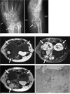
Plain radiographs showed multiple, popcorn-like with some ring- and arc-shaped calcifications with marked soft tissue swelling in left wrist and proximal hand (Fig. 1A). Magnetic resonance imaging showed a large lobulated, hypointense mass on T1-weighted images from the distal metaphysis of the left radius and ulna to the metaphysis of metacarpals, with multifocal bony erosions of the left radius, ulna, carpal bones, and metacarpal bones (Fig. 1B). However, the medullary canals of these bones were not involved. Axial T2-weighted images showed a hyperintense mass with multiple signal-void nodules, suggesting chondroid calcifications on plain radiograph (Fig. 1C). The gadolinium enhanced T1-weighted images showed nodular and septal enhancement in this mass (Fig. 1D). The median nerve, radial vessel, extensor muscles and tendons were compressed and displaced by this mass. Chest CT demonstrated no pulmonary metastasis.
The patient underwent surgical excision of the mass. Upon macroscopic examination, a large lobulated chondroid mass was severely attached to adjacent the bony cortex, muscle, and tendon sheath. Microscopically, this mass had focally infiltrated into adjacent bony cortex, tendon and muscle sheath and it was confirmed as a well-differentiated chondrosarcoma (Fig. 1E).
Discussion
Soft tissue tumors with ring-like or arc-shaped calcification or ossification include extraskeletal chondroma, synovial chondromatosis, mesenchymal chondrosarcoma and synovial chondrosarcoma in differential diagnosis. In our case, a lobulated soft tissue mass encircled the neighboring bones with an equally or similarly dorsal and volar bony erosion along the joint line, suggesting a synovial tumor. By imaging alone, it is not possible to differentiate between primary synovial chondromatosis and synovial chondrosarcoma.
Chondrosarcoma of the synovium is extremely rare. Less than 40 cases have been reported and most of them have occurred in the knee and hip. The clinical features of synovial chondromatosis and synovial chondrosarcoma are same (1). Complaints of joint pain, swelling, and the limitation of motion are common. Local recurrence and soft tissue invasion are found in both conditions (2345).
Radiographically soft tissue swelling is seen in the involved joint and calcified bodies, and marginal bony erosion may be present (2). On MR images, synovial chondrosarcoma is often a lobulated intraarticular soft tissue mass, seen as a hypointense signal on T1-weighted images, turing hyperintense on T2-weighted images (67). When the cartilaginous nodules contain calcification, small nodules of low signal intensity are observed on all pulse sequences. Our case showed clumps of the chondroid calcification on plain radiograph, and a lobular, appearing cartilaginous, tumor mass on MR imaging, which led to diagnosis of a chondroid tumor from synovium. Gadolinium enhanced MR images showed multiple nodular and thickened septal enhancements. Due to the radiological similarities in recurrent primary synovial chondromatosis and malignant transformation to chondrosarcoma (12), the contrast-enhanced images may not be helpful in diagnosing malignant transformations. There was no evidence, such as a true bone marrow invasion to suggest malignant transformation in our case (1).
The majority of patients with malignant transformations of primary synovial chondromatosis have long-standing disease with multiple local recurrences. Distinguishing recurrent disease from malignant transformation can be difficult because the local recurrence of primary synovial chondromatosis can be frequent (678910). The time interval between the primary excision of tumor and the diagnosis of chondrosarcoma ranged from 1 month to 24 years (2456789). Our patient had an initial operation for the palpable mass 8 years ago and the mass was microscopically diagnosed as a synovial chondromatosis. The interval between initial diagnosis and malignant transformation to chondrosarcoma was not definite, but appeared to be approximately 8 years. During the past 8 years she has had 3 operations at the same site. Our patient had no metastatic nodules in the lung.
Many articles emphasized that the microscopic features to establish the histopathological diagnosis of malignant change can also be difficult (235). Histological criteria indicating synovial chondrosarcoma are loss of the typical clustering pattern and marked myxoid change of the matrix, a sheetlike arrangement of the chondrocytes, spindle cell proliferation of the chondrocytes, foci of necrosis, and bone marrow invasion (1).
Treatment of patients with malignant transformation of primary synovial chondromatosis usually requires amputation. Metastasis to the lung is common and is reported in 56% of patients with follow-up (10).
In summary, this is the case of an 82-year-old woman with synovial chondrosarcoma developed from synovial chondromatosis at the same site in the wrist, intercarpal and metacarpocarpal joints. According to the literature, synovial chondrosarcoma in the wrist, intercarpal and carpometacarpal joints is relatively unusual even among synovial chondrosarcomas, therefore we report this case of synovial chondrosarcoma.
 XML Download