

 PDF
PDF ePub
ePub Citation
 Print
Print


Schwannomas are benign, encapsulated and submucosal neural sheath tumors derived from Schwann's cells. Approximately 25 to 45% of all schwannomas present in the head and neck with most occurring in the parapharyngeal space. However, schwannomas of the larynx arising from the aryepiglottic fold, epiglottis, false or true vocal cord are quite rare, accounting for approximately 0.1% of all laryngeal neoplasms (1). It is believed to arise from the internal branch of the superior laryngeal nerve after penetrating the thyroid membrane (2). The signs and symptoms of this lesion include sore throat, odynophagia, dysphagia, dyspnea upon exertion, stridor, hoarseness, and a globus sensation. We report a case of a laryngeal schwannoma arising from both aryepiglottic folds.
Case Report
A 29-year-old female presented with a sore throat that had lasted 3 weeks. The symptoms, including sore throat, dyspnea, dysphagia and foreign body sensation, were aggravated recently. She had no signs of infection, such as fever, chilling, neck swelling or tenderness. The laboratory findings were unremarkable. She was a pregnant woman at 4 weeks' of gestation, and had no smoking history or previous airway infection.
Indirect laryngoscopy revealed a smooth, pedunculated, submucosal mass that was based broadly at the both aryepiglottic folds and the lumen of the hypopharynx was nearly occluded. The vocal cord was not evaluated clearly due to the mass but was grossly normal.
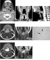
Contrast-enhanced computed tomography (CT) of the neck was performed with a shield protecting the abdomen and pelvis. Axial, reformatted coronal and sagittal images were obtained using 64-channel CT (Brilliance 64; Philips Medical Systems, Cleveland, Ohio). The CT images showed a well-defined, heterogeneously enhancing mass that was intra-luminal protruding and broad-based at the bilateral aryepiglottic folds. The mass showed layered or target-like enhancement on the delayed-phase CT scan after injecting a contrast agent (Figs. 1A-C). On magnetic resonance imaging (MRI), the mass showed slightly hyperintense signal intensity comparing to the adjacent hypoglossal muscles on the T1- and T2-weighted images and heterogeneous enhancement on the gadolinium-enhanced MR images (Figs. 1D-G). The lumen of the hypopharynx and larynx was significantly occluded by the mass.
Antibiotics were administered for 7 days but the symptoms did not improve. Therefore, a laryngomicroscopic excision was carried out under general anesthesia. A 3×2.5 cm whitish, encapsulated mass attached to the bilateral aryepiglottic folds was removed. The mucosal integrity was preserved and the true vocal cords showed normal features after the procedure.
Histologically, the mass was confirmed to be a schwannoma. Two different patterns can usually be recognized, designated Antoni A and B. The type A areas are quite cellular, composed of spindle cells that are often arranged in a palisading manner or in an organoid arrangement (Verocay body). In the type B areas, the tumor cells are separated by abundant edematous fluid that may form cystic space (Fig. 1H).
Discussion
There are two types of benign neurogenic tumors of the larynx involving the aryepiglottic folds; schwannoma and neurofibroma (2). The majority of neurogenic tumors in the larynx originate either from the aryepiglottic fold or false vocal cords, where they bulge into the supraglottic space. Schwannomas are solitary and encapsulated, whereas neurofibromas are non-encapsulated, usually asymptomatic, and may be associated with von Recklinghausen's syndrome. Therefore, a neurofibroma must be considered seriously when neurofibromatosis is observed. In contrast to neurofibromas, which contain a mixture of cell types, schwannomas consist almost exclusively of Schwann cells (3).
Schwannomas of the larynx are rare tumors located at the aryepiglottic fold or false vocal cord. However, the aryepiglottic fold is the most commonly reported site of origin. The most frequently involved nerve is the internal branch of the superior laryngeal nerve. Neurogenic tumors account for 0.1% to 1.5% of all benign laryngeal tumors with schwannomas being more frequent than neurofibromas (2).
Schwannomas may arise at any age (during the 4th and 5th decade of life), and have a female predominance (4). Sanghivi et al. (5) suggested that schwannomas arise from the perineural Schwann cells of a peripheral nerve and grow eccentrically away from the nerve trunk. They are slow-growing and usually solitary.
Patients with a schwannoma of the head and neck region typically present with hoarseness, sore throat, odynophagia, dysphagia, dyspnea, stridor, and/or a foreign body sensation. On laryngoscopy, most lesions appear as a smooth submucosal swelling confined to a false vocal fold or aryepiglottic fold. A small percentage of cases arise on a true vocal fold (4).
Optical microscopy reveals schwannomas to be comprised of a mixture of two growth patterns. In the Antoni A pattern of growth, elongated cells with cytoplasmic processes are arranged in fascicles in areas of moderate to high cellularity with little stromal matrix. The "nuclear-free zones" of the processes that lie between the regions of nuclear palisading are known as Verocay bodies. In the Antoni B pattern of growth, the tumor is less densely cellular with a loose meshwork of cells along with microcysts and myxoid changes. In both areas, the cytology of the individual cells is similar, with elongated cell cytoplasm and regular oval nuclei. Electron microscopy shows basement membrane deposition encasing single cells and long-spacing collagen. Because the lesion displaces the nerve of origin as it grows, silver staining can demonstrate that the axons are largely excluded from the tumor. However, they may become entrapped in the capsule. The Schwann cell origin of these tumors is borne out their S-100 immunoreactivity. A variety of degenerative changes may be found in schwannomas, including nuclear pleomorphisms, xanthomatous changes, and vascular hyalinization. Malignant changes are extremely rare in schwannomas, although local recurrence can follow an incomplete resection (6).
Although comprising a very small proportion of benign laryngeal lesions, schwannomas should be considered in a differential diagnosis. A diagnosis can be made by taking either a fine-needle aspiration or an incisional biopsy of the mass via the endolaryngeal route. However, a diagnosis is difficult without excising the tumor. Confusion between a schwannoma and a neurofibroma might also arise but this should not alter the management plan. CT and/or MRI are valuable techniques for delineating the anatomical extent of the lesion (3).
Laryngeal schwannomas are radioresistant, making a surgical excision the treatment of choice. It is important to remove the tumor completely because of the risk of rapid recurrence that can lead to airway compromise. The size of the tumor dictates the surgical approach. An endoscopic approach is sufficient for smaller tumors, but larger tumors may require an external approach (e.g., lateral pharyngotomy or midline thyrotomy) to achieve complete tumor removal while preserving the laryngeal function and overlying mucosa (7). The patient's prognosis is generally good after complete removal of the tumor.
 XML Download