

 PDF
PDF ePub
ePub Citation
 Print
Print


Inverted Meckel diverticulum is a well-known disorder (12) but difficult to verify when this lesion is encountered in an unusual location. However, this disease can be confirmed when the vitelline artery and vein is identified in the diverticulum (34). We report a case of inverted Meckel diverticulum in an unusual location with the CT demonstration of a vitelline artery and veins, which was confirmed from the pathology.
Case Report
A 28-year-old woman presented with periumbilical pain and hematochezia. This patient reported intermittent periumbilical pain with a 1 year duration. Two months ago earlier, the pain became aggravated and hematochezia developed. Upon admission, the physical examination was normal. The hemoglobin level was 9.5 g/dL (normal=12-18) and the hematocrit level was 29.4% (normal=37-52). The other laboratory tests showed no abnormal findings. Upper gastrointestinal endoscopy and colonoscopy findings were normal.
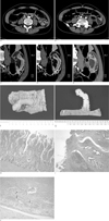
The contrast-enhanced CT scan showed an elongated intraluminal mass with central fat attenuation within the jejunum connected to the mesenteric fat (Figs. 1A-E). The vitelline artery and veins were clearly visualized in the central fatty core ending at the tip of the inverted diverticulum (Figs. 1A-E). These vessels were separate branches that were connected to the superior mesenteric artery and vein.
During surgery nine days later, a small bowel intussusception was observed and a manual reduction of the intussusception was performed. An approximately 9-cm long section of the small bowel containing the mass lesion was resected, and an end-to-end anastomosis was constructed. The segmented small intestine showed an elongated protruding mass covered with pale pink to tan mucosal tissue, measuring 6.5 cm in length and 2.0 cm in diameter (Fig. 1F). The cut section of the diverticular mass showed intersected adipose tissue, muscularis propria lined along the diverticular wall, ulcerated mucosa in the tip of the diverticular mass and vitelline vessels in the proximal portion of the diverticular serosa (Fig. 1G).
The histology examination showed that the diverticulum contained all layers of the intestinal wall. The mucosa was consistent with that normally found in the jejunum (Fig. 1H). The tip of the diverticulum showed focal erosive mucosa and underlying granulation tissue, as well as pancreatic ducts and acinar structure (Fig. 1I). The serosal wall of the diverticulum showed a large artery and venous channels in the central fibroadipose tissue of the diverticulum (Fig. 1J).
Discussion
Most small bowel diverticula are acquired but Meckel diverticulum is the only congenital form. Meckel diverticulum is the true diverticulum that is is located on the antimesenteric border of the small bowel within 40-80 cm from the ileocecal valve (5). However, there are some case reports concerning the presence of congenital jejunal diverticulum (6) or Meckel diverticulum of the proximal jejunum (7).
An understanding the embryologic origin of Meckel diverticulum defines the criteria for this diagnosis as follows: 1) a diverticulum that arises on the antimesenteric border of the small intestine and contains all layers of the intestinal wall; and 2) has a separate blood supply of vitelline vessels originating from the mesenteric vessels but crossing the intestinal wall to supply the diverticulum (7). The mechanism of the proximal origin suggests that Meckel diverticulum might arise anywhere within the boundaries of the midgut. The ultimate position would depend on the relative growth of the intestine proximal and distal to the remnant of the vitelline duct (7). Our case was confirmed to be a congenital diverticulum in view of the presence of a muscularis layer in the gross and microscopic pathology findings and a distal jejunal diverticulum considering the high location in the intraoperative findings, prominent circular folds in the gross pathology findings, and prominent villi and finger-like projections of the mucosa in the microscopic pathology finding.
The structures most commonly confused with Meckel diverticulum are small bowel diverticula and duplications. Small bowel diverticula do not contain all layers of the intestinal wall, do not have a separate blood supply, and are observed along the mesenteric border of the intestine, usually within the leaves of the mesentery. Duplications contain all layers of the intestinal wall, do not have a separate blood supply, and are found along the mesenteric border of the intestine (7).
The angiographic diagnosis of a Meckel diverticulum relies on the observation of a vitellointestinal artery, even though such a vessel may not always be present. The characteristic vitelline artery is reported to be an elongated vessel without anastomosing branches to the ileal branches. This lesion can be diagnosed confidently as an inverted Meckel diverticulum if the vitellointestinal artery and vein can be demonstrated in an inverted diverticulum of an unusual location (34).
The persistence of the right or left vitelline artery and the right or left vitelline vein may be identified in a Meckel diverticulum with the pattern of one artery and one vein (8). However, our case showed one vitelline artery and two vitelline veins.
An inversion of the diverticulum has been suggested to be a primary process that is caused by the inadequate drainage of secretions, heterotopic pancreatic tissue, coproliths, or abnormal peristalsis due to an ulceration of the mucosa (29). Our case had heterotopic pancreatic tissue and areas of ulcerated mucosa at the tip of the diverticulum that could explain the invagination. Although isolated inversion of a diverticulum without intussusception was observed in the CT scan, small bowel intussusception with an inverted Meckel diverticulum was noted at surgery performed nine days later. This is because the inverted diverticulum might serve as a lead point for intussusception.
An inverted Meckel diverticulum appears radiographically as an elongated, smoothly marginated intraluminal mass parallel to the long axis of the bowel. The inverted diverticulum has a bulbous tip with a clublike appearance. The club appears to result from mesenteric fat filling the tip of the inverted sac limited with a serosal surface. A solitary, elongated, clublike mass in the distal ileum depicted on a barium examination is highly suggestive of an inverted Meckel diverticulum (1). The inverted Meckel diverticulum is observed by CT as a central area of fat attenuation surrounded by a thick collar of soft-tissue attenuation. This appearance results pathologically from the entrapment of mesenteric fat within the inverted diverticular sac (1). Ultrasound reveals a target-like mass containing a central focus of increased echogenicity, which is probably related to the core of mesenteric fat in the inverted diverticulum (110).
The differential diagnosis of an elongated, club-shaped mass in the small bowel includes a lipoma, inflammatory fibroid polyp, and other pedunculated polyps. Lipoma is depicted by CT as a lesion with fat attenuation that is not surrounded by a thick collar of soft tissue. An inflammatory fibroid polyp or pedunculated polyps are depicted as a soft-tissue mass without an area of fat attenuation (1).
In conclusion, an inverted Meckel diverticulum produces the characteristic CT findings of an elongated, smoothly marginated intraluminal mass parallel to the long axis of the bowel with a central area of fat attenuation surrounded by a thick collar of soft-tissue attenuation. This diverticulum can be diagnosed confidently as a Meckel diverticulum when a vitellointestinal artery and vein is observed in an unusual location of the diverticulum by CT.
 XML Download