

 PDF
PDF ePub
ePub Citation
 Print
Print


Abstract
Desmoid tumors are rare soft tissue tumors arising from the fascia or from musculoaponeurotic structures. Multifocal desmoid tumors are rare. In patients with Gardner's syndrome or familial polyposis, most cases may occur in the abdominal wall and mesenteries, but are typically only seen after abdominal surgery. A desmoid tumor arising in the terminal ileum is extremely rare. To the best of our knowledge, there is no case report of a sporadic desmoid tumor arising in the terminal ileum and rectus abdominal muscle. We report a case of a sporadic desmoid tumor arising in the terminal ileum and rectus abdominal muscle that presented as a palpable mass of the right upper abdomen in a 38-year-old woman. This finding draws attention to the related findings of previous studies on desmoids tumors.
Figures and Tables
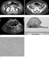
Fig. 1
38-year-old woman with a palpable mass in the right upper abdomen.
A. Enhanced axial CT image shows a well-circumscribed heterogeneously enhancing mass with inner prominent vessels (white arrows) in the right rectus muscle.
B. Enhanced axial CT image shows a well-circumscribed heterogeneously enhancing mass arising from the terminal ileum (white arrows). The mass with radiating strands and small lymph nodes is noted.
C. Transverse sonogram shows a well-defined heterogeneously hypoechoic mass (white arrows) in the right rectus muscle.
D. Gross specimen of desmoid tumor after resection. A partially ill-defined subserosal solid mass with a rubbery pale, tan trabeculated cut surface is noted. There is grossly no evidence of necrosis.
E. Uniform fibroblastic tumor cells are arranged in fascicles and separated by variable amounts of collagen (H/E ×200).
References
1. Singh N, Sharma R, Dorman SA, Dy VC. An unusual presentation of desmoid tumor in the ileum. Am Surg. 2006; 72:821–824.
2. Faria SC, Iyer RB, Rashid A, Ellis L, Whitman GJ. Desmoid tumor of the small bowel and the mesentery. AJR Am J Roentgenol. 2004; 183:118.
3. Lee YI, Lee HK, Hong HS, Kwon KH, Choi DL, Kim JJ. Progression of Desmoid Tumors in Familial Polyposis: A Case Report. J Korean Radiol Soc. 2001; 44:89–92.
4. Kawashima A, Goldman SM, Fishman EK, Kuhlman JE, Onitsuka H, Fukuya T, et al. CT of intraabdominal desmoid tumors: is the tumor different in patients with Gardner's disease? AJR Am J Roentgenol. 1994; 162:339–342.
5. Teo HE, Peh WC, Shek TW. Case 84: desmoid tumor of the abdominal wall. Radiology. 2005; 236:81–84.
6. Einstein DM, Tagliabue JR, Desai RK. Abdominal desmoids: CT findings in 25 patients. AJR Am J Roentgenol. 1991; 157:275–279.
7. Kim MJ, Park KJ, Sun JS, Kim JH, Choi H. Intrathoracic Desmoid Tumor: A Case Report and Radiological Evaluation. J Korean Radiol Soc. 2007; 57:31–35.
8. Kreuzberg B, Koudelova J, Ferda J, Treska V, Spidlen V, Mukensnabl P. Diagnostic problems of abdominal desmoid tumors in various locations. Eur J Radiol. 2007; 62:180–185.
9. Newman CA, Reuther WL 3rd, Wakabayashi MN, Payette MM, Plavsic BM. Gastrointestinal case of the day. Gardner syndrome. Radiographics. 1999; 19:546–548.
10. Casillas J, Sais GJ, Greve JL, Iparraguirre MC, Morillo G. Imaging of intra- and extraabdominal desmoid tumors. Radiographics. 1991; 11:959–968.
 XML Download