

 PDF
PDF ePub
ePub Citation
 Print
Print


Abstract
Purpose
To establish recommendation criteria for the determination of patient dose for a plain chest radiography examination (PCR) by surveying the examination protocols for the performance of PCR, determining patient doses, and analyzing the relationship between these protocols and patient dose.
Materials and Methods
We surveyed the examination protocols for the PCR of 95 radiography units in 82 institutes. We measured the patient dose as the surface entrance dose (SED) in these unites using an anthropomorphic phantom. We ultimately compared the results of the survey against several significant criteria.
Results
The protocol for performing a PCR in Korea was 111 ± 17 kVp and 7.0 ± 6.7 mAs. The mean and median SED were 0.30 ± 0.27 mGy and 0.22 mGy, respectively. The third quartile value was 0.34 mGy, while the variability between the types of institutes and the average values of SED were 0.27 ± 0.17 mGy, 0.28 ± 0.29 mGy, and 0.34 ± 0.31 mGy for primary, secondary, and tertiary hospitals, respectively. The types of units include the average values of SED, which were 0.18 ± 0.11 mGy, 0.37 ± 0.28 mGy, and 0.32 ± 0.31 mGy for film/screen, CT, and digital radiography systems, respectively.
Figures and Tables
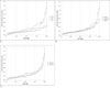
Fig. 3
Entrance surface dose value plots for the included units as ascending order.
A. Comparison between film/screen, storage phosphor, and flat panel digital radiography systems
B. Comparison between primary, secondary, and tertiary hospitals
C. Comparison between low (< 100 kVp) and high (≥ 100 kVp) tube voltage conditions


References
1. Turai I, Veress K, Gunalp B, Souchkevitch G. Major radiation exposure. N Engl J Med. 2002; 347:944–947. author reply 944-947.
2. ICRP. Recommendations of the International Commission on Radiological Protection, Publication 60. Oxford: Pregamon Press;1990.
3. Contento G, Malisan MR, Padovani R, Maccia C, Wall BF, Shrimpton PC. A comparison of diagnostic radiology practice and patient exposure in Britain, France and Italy. Br J Radiol. 1988; 61:143–152.
4. Shrimpton PC, Wall BF, Jones DG, Fisher ES, Hillier MC, Kendall GM, et al. Doses to patients from routine diagnostic X-ray examinations in England. Br J Radiol. 1986; 59:749–758.
5. Warren-Forward HM, Millar JS. Optimization of radiographic technique for chest radiography. Br J Radiol. 1995; 68:1221–1229.
6. Physical Sciences in Medicine NRPBatCoR. National protocol for patient dose measurements in diagnostic radiology. Chilton, Didcot: NRPB;1992.
7. 97/43/Euratom Cd. Health protection of individuals against the dangers of ionizing radiation in relation to medical exposure, and repealing Directive 84/466/Euratom: The European Commission. 1997.
8. Wall BF, Hart D. Revised radiation doses for typical X-ray examinations. Report on a recent review of doses to patients from medical X-ray examinations in the UK by NRPB. National Radiological Protection Board. Br J Radiol. 1997; 70:437–439.
9. Hosch WP, Fink C, Radeleff B, kampschulte A, Kauffmann GW, Hansmann J. Radiation dose reduction in chest radiography using a flat-panel amorphous silicon detector. Clin Radiol. 2002; 57:902–907.
10. Bacher K, Smeets P, Bonnarens K, De Hauwere A, Verstraete K, Thierens H. Dose reduction in patients undergoing chest imaging: digital amorphous silicon flat-panel detector radiography versus conventional film-screen radiography and phosphor-based computed radiography. AJR Am J Roentgenol. 2003; 181:923–929.
11. Ganten M, Radeleff B, Kampschulte A, Daniels MD, Kauffmann GW, Hansmann J. Comparing image quality of flat-panel chest radiography with storage phosphor radiography and film-screen radiography. AJR Am J Roentgenol. 2003; 181:171–176.
12. Warren-Forward H, Arthur L, Hobson L, Skinner R, Watts A, Clapham K, et al. An assessment of exposure indices in computed radiography for the posterior-anterior chest and the lateral lumbar spine. Br J Radiol. 2007; 80:26–31.
 XML Download