

 PDF
PDF ePub
ePub Citation
 Print
Print


The combination of positron emission tomography (PET) and computed tomography (CT) are increasingly used for oncologic imaging. In particular, positron emission tomography with fluorine-18 fluorodeoxyglucose (18F-FDG PET/CT) is suggested as a more useful modality for accurate, non-invasive imaging in predicting the prognosis and staging of breast cancer (1234). The combination of positron emission tomography (PET) and computed tomography (CT) provides functional metabolic information (PET) and morphologic information. 18F-FDG PET/CT has been evaluated for primary breast cancer detection and diagnosis, staging of locoregional and distant sites, and monitoring the response to therapy in previous studies (567). Although 18F-FDG PET/CT is widely recognized as a useful diagnostic tool, it produces false-negative results in 12% of cancer cases (8).
The aim of this study was to identify radio-clinicopathologic factors that predict false negative FDG uptake results in breast cancer on 18F-FDG PET/CT.
Materials and Methods
Patients
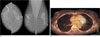
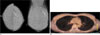
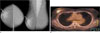
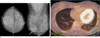
We retrospectively reviewed a total of 140 breast cancers in 140 patients (mean age, 51.3 years: range, 28-86 years) from May 2007 to January 2008. All patients were histologically or cytologically confirmed as having breast cancer before undergoing 18F-FDG PET/CT. All patients were examined with 18F-FDG PET/CT for staging of breast cancer. This retrospective study was approved by the ethics review committee and informed consent was obtained from all patients. According to the results of 18F-FDG PET/CT, the patients were divided into two groups: Group 1 consisted of 20 patients who had negative results for the primary mass. Group 2 consisted of 120 patients who had positive results for the primary mass. The radio-clinico-pathologic factors including patient's age, tumor size, estrogen receptor (ER), progesterone receptor (PR), C-erb-B2, types of pathology and mammography findings, the inclusion of the mass (present, not present), clustered calcification (present, not present), and breast parenchyma composition (fatty breast, scattered fibroglandular tissue, heterogeneous fibroglandular tissue, dense breast) of group 1 and group 2 were retrospectively reviewed.
Mammography
A bilateral mammography (MAMMOMAT NovationDR, Siemens Medical Solutions, Forchheim, Germany), including routine craniocaudal and mediolateral oblique views of the breasts, was performed. Findings were recorded prospectively according to BI-RADS by two radiologists who had 2 and 10 years of experience in performing mammographies. A mammography was performed at least 4 weeks before the other studies.
PET Scanning
18F-FDG PET/CT was performed with a dedicated PET/CT scanner (Gemini, Philips Medical System, Milpitas, CA, USA), consisting of a germanium oxyorthosilicate full-ring PET scanner and a dual slice helical CT scanner. Standard patient preparation included at least 8 hours of fasting to attain a serum glucose level of less than 120 mg/dL before 18F-FDG administration. PET/CT imaging was performed 60 minutes after the injection of 4.5 MBq/Kg of 18F-FDG. At 60 minutes after administering 18F-FDG, low-dose CT (30 mAs, 120kV) covering an area from the base of the skull to the proximal thighs was performed for the purpose of attenuation correction and precise anatomical localization. Therefore, an emission scan was conducted in 3-dimensional mode. The emission scan time per bed position was 3 minutes; a total of 9 bed positions were acquired. PET data were obtained using a high resolution whole body scanner with an axial field of view of 18 cm. The average total PET/CT examination time was 30 minutes. After scatter and decay correction, PET data were reconstructed iteratively with attenuation correction and reoriented in axial, saggital, and coronal slices. The row action maximum-likelihood algorithm was used for 3-dimensional reconstruction.
For positive findings on the 18F-FDG PET/CT image, we relied on a visual focus of the PET image (a well-defined focus with uptake clearly greater than the surrounding background) and excluded the underlying morphologic CT information. A cut-off maximum standardized uptake value of 2.5 was applied to discriminate the positive and negative PET results. All 18F-FDG PET/CT images were directly reviewed on a computer workstation.
Histopathological and Immunohistochemical Study
Formalin-fixed, paraffin-embedded sections of the resected mass were stained with hematoxylin-eosin (HE) and analyzed. Immunohistochemical analyses for the estrogen receptor (ER), progesterone receptor (PR), and c-erb-B2 (proto-oncogene) were performed using specific monoclonal antibodies.
Statistical Analysis
Univariate and multivariate analyses were used for comparison of the two groups.
For the univariate analysis, age and size were compared using the Mann-Whitney U test; histologic results, mammography findings, ER, PR, and C-erb-B2 were compared by the Chi-squared test. P-values less than 0.05 were considered statistically significant.
For the statistical analysis, histologic results were divided into ductal carcinoma in situ and lobular carcinoma in situ versus invasive ductal carcinoma, invasive papillary carcinoma and mucinous carcinoma. Also, the breast composition among the mammography findings was divided into fatty breast and scattered fibroglandular tissue versus heterogeneous fibroglandular tissue and dense breast.
For the multivariate analysis, all factors were compared by stepwise logistic regression analysis.
Results
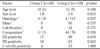
The mean size of the masses were 1.8 cm (range, 1.2-3.5 cm) in group 1 and 2.4 cm (range, 1.5-5.4 cm) in group 2. The mean maximum standardized uptake values were 0.5 (range, 0-1.7) in group 1 and 6.1 (range, 2.6-9.6) in group 2. The other results are summarized in Table 1. Among all parameters, estrogen receptor positivity (p = 0.019), progesterone receptor positivity (p = 0.01), carcinoma in situ (p = 0.037), and the size of the mass (p = 0.012) were analyzed by Mann-Whitney test or Chi-square test and found to show a statistical difference between groups 1 and 2. A stepwise logistic regression analysis showed that estrogen receptor positivity (odds ratio, 5.623; 95% confidence interval: 1.100, 28.746; p = 0.021) and carcinoma in situ (odds ratio, 6.900; 95% confidence interval: 1.151, 41.361; p = 0.026) were significant clinico-pathology variables associated with a negative finding for 18F-FDG PET/CT in the diagnosis of primary breast cancer (Table 2).
Discussion
Currently, a whole body CT, bone scintigraphy, and breast magnetic resonance imaging are used for the initial staging of the tumor and the detection of distant metastasis. 18F-FDG PET/CT is proposed as a single method that can replace these methods (9). 18F-FDG PET/CT provides functional, metabolic information, and morphologic information. However, Samson et al. reported that false negative results occurred in 12% of breast cancers (8).
Our results showed that the estrogen receptor positivity and carcinoma in situ were significant clinico-pathology variables associated with the negative findings of 18F-FDG PET/CT in the diagnosis of primary breast cancer. Therefore, in cases of estrogen receptor positivity and carcinoma in situ tumors, there is a high possibility that the primary mass and metastatic mass in the contralateral breast are not depicted on 18F-FDG PET/CT for stage workup.
Kumar et al. (10) reviewed 85 breast cancers and demonstrated that both tumor sizes of less than 10 mm and low tumor grade were significant predictors of a false-negative 18F-FDG PET/CT result. However, to our knowledge, there has been no report that estrogen receptor positivity is associated with a negative 18F-FDG PET/CT finding in the diagnosis of breast cancer.
Generally, estrogen receptor positive tumors are known to be less aggressive. Therefore, we considered that our result was attributed to the fact that in less aggressive tumors, glucose metabolism more slowly accelerates to meet the energy demand for tumor growth. Despite the fact that the estrogen receptor is still not completely understood, we hope that the knowledge gained from this study contributed to explaining 18F-FDG uptake rates.
Despite its several advantages, it is still questionable whether 18F-FDG PET/CT is useful for the stage workup of a tumor showing no 18F-FDG uptake. In fact, the current National Comprehensive Cancer Network practice guidelines recommend that routine chest imaging (chest radiography) should only be performed on patients with clinical stage I breast cancer. In patients with node-positive stage II and stage III disease, imaging typically consists of bone scanning and contrast-enhanced chest or abdominal CT. 18F-FDG PET/CT is recommended as an option for patients with either recurrent or stage IV disease, which in this setting, has been shown to be both sensitive and specific for metastases (111213).
There is widespread agreement that whole-body 18F-FDG PET/CT does not have a clinical role in detecting primary breast cancer, nor is it an alternative to histologic sampling to establish or exclude primary breast cancer because of the well-documented inability of 18F-FDG PET/CT to consistently demonstrate small and low-grade lesions (14). Therefore, other imaging modalities are required for the precise detection of breast cancer and metastasis evaluation. In some studies, the sensitivity and specificity of MR imaging were higher for BRCA mutation carriers (1516). In another study, a relatively high number of cancers (13 of 33 [39%]) were only visible on MR imaging, other than US and mammography (17).
Our study had three limitations: First, although the reference standard in this study was based on results from the combined analysis of detailed standardized pathologic reports and imaging studies, inaccuracies might have been introduced because of the retrospective nature of this study. Second, we did not include ultrasonography findings in the radio-clinico-pathologic factors, which are important diagnostic factors. Third, we considered the mass on the CT scan, without 18F-FDG uptake, as a negative finding. However, our study focused on 18F-FDG uptake, not morphologic information.
In conclusion, carcinoma in situ and ER positivity were significantly correlated with false negative FDG uptake in breast cancer on 18F-FDG PET/CT.

 XML Download