

 PDF
PDF ePub
ePub Citation
 Print
Print


Abstract
Purpose
To evaluate the diagnostic value of oblique coronal reconstructed CT images to determine the local invasion of advanced gastric cancer (AGC).
Materials and Methods
Thirty-four consecutive patients, who were suspected to have locally invasive advanced gastric cancer (more than T3 stage) on a preoperative MDCT scan and underwent a diagnostic or curative laparotomy, were enrolled in this study. Two reviewers performed an independent blind review of three series of MDCT images in random order; axial (AXI), conventional coronal (CCI), and oblique coronal (OCI) (parallel to long axis of gastric body and pancreas) images. In assessing the local invasion, the reader's confidence for the local invasion of AGC was graded using a five point scale (1 = definitely negative, 5 = definitely positive: T4). With surgical findings and histopathological proofs as reference standards, the diagnostic performance of the three different plans of CT images was employed for the verification of local invasion of AGC on a preoperative CT scan using the receiver operating characteristic (ROC) method. Agreements between the two reviewers were analyzed using weighted kappa statistics.
Results
In 19 out of 34 patients, local invasion was confirmed surgically or histopathologically (13 pancreas invasion, 6 liver invasion, 4 major vascular invasion, 3 colon and mesocolon invasion, and 2 spleen invasion). The diagnostic performance of OCI was superior to AXI or CCI in the local invasion of AGC. The differences in the area under the curve of AXI (0.770 ± 0.087, 0.700 ± 0.094), CCI (0.884 ± 0.058, 0.958 ± 0.038), and OCI (0.954 ± 0.050, 0.956 ± 0.049), were statistically significant for both reviewers. Inter-observer agreement was excellent for OCI (κ= .973), which was greater than CCI (κ= .839), and AXI (κ= .763).
Figures and Tables
Fig. 1
Oblique coronal image.
Oblique coronal images were obtained by images parallel to the long axis of the gastric body and the pancreas
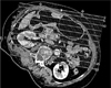
Fig. 2
MDCT of Advanced Gastric Cancer without Local Invasion in 43-year-old female.
A, B. Irregular gastric wall-thickened mass (M), suggestive of advance gastric tumor, is in contact (arrows) with pancreas on axial (A) and coronal (B) images.
C. On oblique coronal image, mass is separated from pancreas by intact fat plane (narrow arrows). At surgical field, there was no direct invasion into the pancreas.
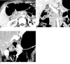
Fig. 3
MDCT of Advanced Gastric Cancer without Local Invasion in 39-year-old female.
A. Axial image shows fat plane was effaced with subtle irregular interface (arrowheads) between gastric tumor(M) and pancreas on axial image.
B, C. Coronal (B) and oblique coronal (C) images show distinct and smooth interface (arrows) between gastric tumor (M) and pancreas, without compression. At surgery, there was no direct invasion into the pancreas.
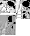
Fig. 4
MDCT of Advanced Gastric Cancer with Local Invasion in 66-year-old male.
A, B. Irregular gastric wall-thickened mass, suggestive of advance gastric tumor, shows subtle irregular interface (arrows) between the tumor mass (M) and the left hepatic lobe on axial (A) and coronal (B) images. But, direct invasion of the liver by the tumor is not clear on given CT images.
C. On oblique coronal image, however, hepatic invasion (arrows) is shown more clearly.
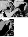
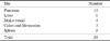
Table 2
Comparison of Local Invasion of Advanced Gastric Cancer Between Pathologic and CT Staging with Axial Imaging

Table 3
Comparison of Local Invasion of Advanced Gastric Cancer Between Pathologic and CT Staging with Coronal Imaging

Table 4
Comparison of Local Invasion of Advanced Gastric Cancer between Pathologic and CT Staging with Oblique Coronal Imaging


References
1. National Cancer Center. Cancer Facts & Figures 2009. Goyang: National Cancer Center;2009. p. 24–25.
2. Shirakawa T, Fukuda K, Tada S. New method for evaluation of perigastric invasion of gastric cancer by right lateral position CT. Eur Radiol. 1996; 6:358–361.
3. Kumano S, Murakami T, Kim T, Hori M, Iannaccone R, Nakata S, et al. T staging of gastric cancer: role of multi-detector row CT. Radiology. 2005; 237:961–966.
4. Botet JF, Lightdale CJ, Zauber AG, Gerdes H, Winawer SJ, Urmacher C, et al. Preoperative staging of gastric cancer: comparison of endoscopic US and dynamic CT. Radiology. 1991; 181:426–432.
5. Hur J, Park MS, Lee JH, Lim JS, Yu JS, Hong YJ, et al. Diagnostic accuracy of multidetector row computed tomography in T- and N staging of gastric cancer with histopathologic correlation. J Comput Assist Tomogr. 2006; 30:372–377.
6. Fukuya T, Honda H, Kaneko K, Kuroiwa T, Yoshimitsu K, Irie H, et al. Efficacy of helical CT in T-staging of gastric cancer. J Comput Assist Tomogr. 1997; 21:73–81.
7. Koo YB, Kim S, Lee JW, Kim SJ, Choo KS, Lee TH, et al. The usefulness of multiplanar reconstruction images in preoperative T-staging of advanced gastric cancer. J Korean Radiol Soc. 2004; 51:241–248.
8. Kim YH, Lee KH, Park SH, Kim HH, Hahn S, Park DJ, et al. Staging of T3 and T4 gastric carcinoma with multidetector CT: added value of multiplanar reformations for prediction of adjacent organ invasion. Radiology. 2009; 250:767–775.
9. Chen CY, Hsu JS, Wu DC, Kang WY, Hsieh JS, Jaw TS, et al. Gastric cancer: preoperative local staging with 3D multi-detector row CT--correlation with surgical and histopathologic results. Radiology. 2007; 242:472–482.
10. Yoo HM, Cho JS, Shin KS, Sung JK, Jeong HY, Noh SM, et al. Usefulness of Helical CT for the preoperative evaluation of small advanced gastric cancer mimicking as early gastric cancer at endoscopy. J Korean Radiol Soc. 2005; 52:385–393.
11. Fleming ID, Cooper JS, Henson DE. Cancer staging manual. 5th ed. Philadelphia: Lippincott-Raven;1997. p. 71–77.
12. Kim YN, Choi D, Kim SH, Kim MJ, Lee SJ, Lee WJ, et al. Gastric cancer staging at isotropic MDCT including coronal and sagittal MPR images: endoscopically diagnosed early vs. advanced gastric cancer. Abdom Imaging. 2009; 34:26–34.
13. Matsushita M, Oi H, Murakami T, Takata N, Kim T, Kishimoto H, et al. Extraserosal invasion in advanced gastric cancer: evaluation with MR imaging. Radiology. 1994; 192:87–91.
 XML Download