

 PDF
PDF ePub
ePub Citation
 Print
Print


Abstract
Purpose
To evaluate the imaging findings of uterus didelphys with obstructed hemivagina and ipsilateral renal agenesis.
Materials and Methods
From March 2003 to December 2008, five patients with uterus didelphys with obstructed hemivagina and ipsilateral renal agenesis were evaluated as part of this study. We retrospectively reviewed the CT, ultrasound, and MRI findings as well as the medical records of each patient.
Results
The patients initially underwent an imaging study for abdominal pain (n=3), recurrent vaginal bleeding (n=1), and prenatal evaluation (n=1). Of the five patients that underwent US, four had hematocolpos and two of them had hematometra. Moreover, three patients underwent a CT examination. The MR examination of four patients revealed hematocolpos (n=3), hematometra (n=1), and a tubular structure resembling an ectopic ureter (n=2). The gynecologic examination of a patient without hematocolpos revealed a pinpoint hole in the vaginal septum. Two of four patients with hematocolpos underwent a vaginal septectomy, which resulted in an improvement of the symptoms.
Conclusion
The most common finding of patients with uterus didelphys with obstructed hemivagina and ipsilateral renal agenesis is vaginal fluid collection. Hematometra is not a consistent finding and can be transient according to the menstrual cycle. MR is the most useful imaging modality for the diagnosis of an ectopic ureter.
Figures and Tables
Fig. 1
Case 1, A 10-year-old girl with abdominal pain.
A. Transabdominal US image shows didelphic uterus and low echoic fluid collection in right uterine cavity (black arrows). Left side horn shows no abnormal fluid retention (white arrows).
B. Axial image of vagina shows dilated right hemivagina with internal heterogeneous hypoechoic lesion with fluid collection suggesting hematoma (arrows).
C, D. T1(C) and T2 (D) weight axial MR image shows didephic uterus and dilated right hemiuterus containing high signal intensity fluid (arrows).
E. T2 weighted axial MR image shows dilated right hemivagina filled with heterogeneous high signal intensity fluid collection, indicating hematocolpos (arrows).
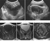
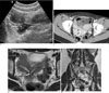
Fig. 2
Case 2, A 16-year-old girl with abdominal pain during menstrual periods.
A. Longitudinal US scan shows dilated right hemivagina with low echogenic fluid (arrows).
B. Axial contrast-enhanced CT scan shows dilated right vaginal canal (arrows) and fat infiltration in pelvic cavity.
C, D. Axial (C) and coronal (D) T2 weighted MR images show didephic uterus, separated cervix, double vagina with fluid collection in right vaginal canal (white arrows). High signal intensity tubal structure is seen on right lateral aspect of uterus suspecting ectopic right ureter (thick arrows). Right renal agenesis is suggested on coronal image.
References
1. Orazi C, Lucchetti MC, Schingo PM, Marchetti P, Ferro F. Herlyn-Werner-Wunderlich syndrome: uterus didelphys, blind hemivagina and ipsilateral renal agenesis. Sonographic and MR findings in 11 cases. Pediatr Radiol. 2007; 37:657–665.
2. Gholoum S, Puligandla PS, Hui T, Su W, Quiros E, Laberge JM. Management and outcome of patients with combined vaginal septum, bifid uterus, and ipsilateral renal agenesis (Herlyn-Werner-Wunderlich syndrome). J Pediatr Surg. 2006; 41:987–992.
3. Stassart JP, Nagel TC, Prem KA, Phipps WR. Uterus didelphys, obstructed hemivagina, and ipsilateral renal agenesis: the University of Minnesota experience. Fertil Steril. 1992; 57:756–761.
4. O’Neill MJ, Yoder IC, Connolly SA, Mueller PR. Imaging evaluation and classification of developmental anomalies of the female reproductive system with an emphasis on MR Imaging. AJR Am J Roentgenol. 1999; 173:407–416.
5. Troiano RN, McCarthy SM. Mu ¨llerian duct anomalies: imaging and clinical issues. Radiology. 2004; 233:19–34.
6. Purslow CE. A case of unilateral haematocolpos, haematomethra and haematosalpinx. J Obstet Gynaecol Br Emp. 1922; 29:643.
7. Vercellini P, Daguati R, Somigliana E, Vigano . P, Lanzani A, Fedele L. Asymmetric lateral distribution of obstructed hemivagina and renal agenesis in women with uterus didelphys: institutional case series and a systematic literature review. Fertil Steril. 2007; 87:719–724.
8. Tanaka YO, Kurosaki Y, Kobayashi T, Eguchi N, Mori K, Satoh Y, et al. Uterus didelphys associated with obstructed hemivagina and ipsilateral renal agenesis: MR findings in seven cases. Abdom Imaging. 1998; 23:437–441.
 XML Download