

 PDF
PDF ePub
ePub Citation
 Print
Print


Peripheral T-cell lymphoma (PTCL) is a subtype of non-Hodgkin's lymphoma (NHL) and PTCL is quite rare in western countries: this accounting for 10 to 15% of all the case of NHL in Europe (1). Although PTCL may involve many organs, including the sino-nasal cavity and airway, intestinal tract, skin, lymph nodes and liver, it rarely involves the lung, and there are few radiological descriptions to guide making the proper diagnosis (2). We report here on a case of PTCL, unspecified (PTCL-U) that which did involve the lung parenchyma and mediastinal lymph nodes, and we present the clinical manifestations and pathological findings.
Case Report
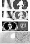
A 39-year-old man was admitted with a mild fever and cough of 3 days duration. The laboratory findings were an erythrocyte sedimentation rate (ESR) of 24 mm/hr, a C-reactive protein (CRP) level of 77 mg/L, the cytoplasmic antineutrophil cytoplasmic antibody (cANCA) was negative and the perinuclear anti-neutrophilic cytoplasmic antibody (pANCA) was negative. The chest radiography on admission revealed a cavitary lesion in the left middle lung zone (Fig. 1A). The chest CT image showed a thin-walled cavitary lesion in the superior segment of the left lower lobe (Fig. 1B). The upper level of the chest CT image, through the carina, revealed multiple lymphadenopathies in the mediastinum. The patient was treated with antibiotics and then he was discharged after the fever and cough had subsided.
About 2 months later, the patient was readmitted to our hospital after 2 days of high fever. The laboratory findings were a white blood cell count of 14, 500/uL, a CRP level of 103 mg/L, the C-ANCA and P-ANCA were negative and the sputum acid-fast bacilli was negative. The antibody titer for Epstein-Barr virus was not elevated. Chest radiography showed an ill-defined consolidation in the left middle lung field and a prominent left hilum (Fig. 1C). The follow-up chest CT images showed a thick-walled cavity in the left lower lobe that was more enlarged than that seen on the previous chest CT scan (Fig. 1D). The mediastinal window images of the chest CT scan revealed multiple lymphadenopathies with poorly defined margins in the mediastinum and an irregular diffuse infiltration in the mediastinal fat (Fig. 1E).
The consolidation with the cavitary lesion was removed by performing video-assisted thoracoscopic surgery. Macroscopically, the cut specimen showed parenchymal hemorrhage and necrosis with a cavitary lesion (Fig. 1F). Microscopically, the specimen showed a cavitary lesion and dilated vessels with surrounding hemorrhagic necrosis that showed angiocentric, pleomorphic, atypical lymphoid cells on hematoxylin and eosin staining (Fig. 1G). Immunohistochemical staining showed positive reactions for cluster of differentiation 3 (CD 3), which is a T cell marker, and for T cell intracytoplasmic antigen (TIA), which is a cytotoxic granule-associated protein, in the pleomorphic atypical lymphoid cells. Staining for CD56, a natural killer/T (NK/T) cell marker, and CD 20, a B cell marker, was negative (Fig. 1H).
Discussion
Peripheral T-cell lymphoma of the unspecified type accounts for 4 to 11% of all non-Hodgkin lymphomas, and pulmonary involvement occurs in 6 to 10% of all patients with PTCL-U (34). The median age of onset for PTCL-U is reported to be in the seventh decade (45). This lymphoma usually takes an aggressive course, with relapses being more common than for B-cell lymphomas. Most patients with PTCL-U present with nodal involvement, but the disease can also involve extranodal sites, including the liver, bone marrow, spleen, gastrointestinal tract and skin (2).
On CT scans, the pulmonary manifestations of lymphoma generally appear as multiple nodules or masslike areas of consolidation with pleural effusion and lymph node enlargement being visible in 42% and 35% of patients, respectively, and without ground glass opacities or reticular opacities (6). Yet pulmonary involvement of PTCL-U has rarely been reported. Lee et al. (2) reported that generalized lymphadenopathies were the most common findings for this lymphoma. The CT findings of lymph node involvement in the neck of PTCL patients were reported as central necrosis, an ill-defined margin and heterogeneous enhancement (7). In our case, the lymph nodes lost their defined margins and there was an irregular diffuse infiltration in the mediastinal fat. Mavi et al. (8) reported on the pulmonary involvement of PTCL with multiple cavitary lesions in both lungs. Our case also showed a thick-walled cavitary lesion on the CT images. This cavitary lesion in the lung pathologically represented necrosis, which is a rare finding of lymphoma (9). However, AIDS-related pulmonary lymphoma usually showed multiple nodules that may be cavitated (10).
Pathologically, the peripheral T-cell lymphomas, unspecified (PTCL-U) that do not match one of the defined entities of PTCL show a variety of cellular morphologies, including medium-sized cells, mixed medium and large cells, large cells and lymphoepithelioid cells (23). Immunohistochemical staining of PTCL-U produces positive reactions for CD 3, a T cell marker, and TIA, a cytotoxic granule-associated protein in the pleomorphic atypical lymphoid cells, but there are negative reactions for CD56, a marker for NK/T cells, and CD 20, a B cell marker. Peripheral T-cell lymphoma, unspecified, also gives a negative reaction for Epstein-Barr virus (EBV), and extranodal NK/T cell marker (310).
In summary, we reported here on a case of peripheral T-cell lymphoma, unspecified, with pulmonary parenchymal involvement that presented as a thickwalled cavitary lesion. Lymphadenopathies with poorly defined margins and a surrounding infiltration in the mediastinal fat may also reflect the severity of the underlying disease, as was noted in this case of peripheral T cell lymphoma, unspecified.
 XML Download