

 PDF
PDF ePub
ePub Citation
 Print
Print


Abstract
A very rare case of multi-organ volvulus, serially involving the spleen, colon and stomach, is presented in a 24-year-old female patient with Down syndrome. This case is of interest because of the three different types of volvulus or torsion that occurred serially over thirteen years in the same patient. We report the imaging findings and suggest possible pathogenesis by a review of the operation record and literature.
Figures and Tables
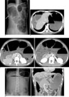
Fig. 1
A. Supine simple abdomen shows markedly gaseous distension of the colon. Subsequent CT showed this was whole colon except the part of rectosigmoid colon. Air filled appendix is shown (arrow).
B-D. Contrast-enhanced axial CT scans. B. At the level of the liver dome, CT scan shows displaced stomach on right side of the liver and markedly distended colon with poorly enhanced wall. C. At the level of SMA origin, CT scan shows markedly distended colon with poorly enhanced wall. Note gas distended appendix on the right side. D. At the level 2 cm below C, CT scan shows totally obliterated SMA (arrow).
E. Erect simple abdomen shows severely distended stomach with air-fluid level.
F. Coronal reformatted CT reveals that the esophagogastric junction (short arrow) is below the pylorus (long arrows). The CT was undertaken after emergent gastric decompression through the nasogastric tube. Note the nasogastric tube within the esophagogastric junction.
References
1. Lehance CW, Gold DM. Colonic volvulus - an old problem with a new twist. Colorectal Dis. 2009; 11:882–883.
2. Learmonth JR. Elongation and dilatation of the colon. Br Med J. 1937; 2:154–156.
3. Bakir B, Poyanli A, Yekeler E, Acunas G. Acute torsion of a wandering spleen: imaging findings. Abdom Imaging. 2004; 29:707–709.
4. Herman TE, Siegel MJ. CT of acute splenic torsion in children with wandering spleen. AJR Am J Roentgenol. 1991; 156:151–153.
5. Berrocal T, Lamas M, Gutieerrez J, Torres I, Prieto C, del Hoyo ML. Congenital anomalies of the small intestine, colon, and rectum. Radiographics. 1999; 19:1219–1236.
6. Cherukupalli C, Khaneja S, Bankulla P, Schein M. CT diagnosis of acute gastric vovulus. Dig Surg. 2003; 20:497–499.
7. Kotobi H, Auber F, Otta E, Meyer N, Audry G, Helardot PG. Acute mesenteroaxial gastric volvulus and congenital diaphragmatic hernia. Pediatr Surg Int. 2005; 21:674–676.
 XML Download