

 PDF
PDF ePub
ePub Citation
 Print
Print


Abstract
Novel influenza A (H1N1) infection is a highly infectious disease, which has been rapidly spreading worldwide since it was first documented in March of 2009 in Mexico. We experienced and report two cases of Influenza A (H1N1) pneumonia, accompanied by chest radiographic and CT findings. The chest radiographs revealed diffuse haziness and extensive airspace consolidation, whereas the CT scans demonstrated multifocal areas of ground glass opacity and airspace consolidation with a CT halo sign.
Figures and Tables
Fig. 1
A 52-year-old previously healthy woman, with no recent history of travel or contact with ill-individuals.
A. An anteroposterior chest radiograph shows both parahilar haziness and suspicious consolidation in the right suprahilar region.
B-D. HRCT scans show multifocal areas of peribronchial and subpleural ground-glass opacification and some consolidation. Note smooth interlobular septal thickening and minimal bilateral pleural effusion.
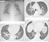
Fig. 2
A 73-year-old asthmatic woman, with a recent history of travel to United States.
A, B. HRCT scans obtained from outside hospital show multifocal areas of consolidation with surrounding ground-glass opacification in both lungs.
C. Chest radiograph at the time of admission to our hospital shows extensive bilateral airspace consolidation, which suggests multifocal pneumonia combined with ARDS. A small amount of pleural effusion is noted in the right upper lateral portion.
D. Follow-up chest radiographs four days later shows persistent bilateral pulmonary opacity with an increased amount of pleural effusion. The patient eventually expired due to respiratory failure.

References
1. Perez-Padilla R, de la Rosa-Zamboni D, Ponce de Leon S, Hernandez M, Quiñones-Falconi F, Bautista E, et al. Pneumonia and respiratory failure from swine-origin influenza A (H1N1) in Mexico. N Engl J Med. 2009; 361(13):680–689.
2. Baker MG, Wilson N, Huang QS, Paine S, Lopez L, Bandaranayake D, et al. Pandemic influenza A (H1N1)v in New Zealand: the experience from April to August 2009. Euro Surveill. 2009; 14(34):pii=19319.
3. Hahne S, Donker T, Meijer A, Timen A, van Steenbergen J, Osterhaus A, et al. Epidemiology and control of influenza A (H1N1)v in the Netherlands: the first 115 cases. Euro Surveill. 2009; 14(27):pii=19267.
4. Gilsdorf A, Poggensee G. Working Group Pandemic Influenza A (H1N1)v. Influenza (H1N1)v in Germany: the first 10,000 cases. Euro Surveill. 2009; 14(34):pii=19318.
5. Kim EA, Lee KS, Primack SL, Yoon HK, Byun HS, Kim TS, et al. Viral pneumonias in adults: radiologic and pathologic findings. Radiographics. 2002; 22:S137–S149.
6. Lee CW, Seo JB, Song JW, Lee HJ, Lee JS, Kim MY, et al. Pulmonary complication of novel influenza A (H1N1) infection: imaging features in two patients. Korean J Radiol. 2009; 10:531–534.
7. Agarwal PP, Cinti S, Kazerooni EA. Chest Radiographic and CT Findings in Novel Swine-Origin Influenza A (H1N1) Virus (S-OIV) Infection. AJR Am J Roentgenol. 2009; 193:1488–1493.
 XML Download