

 PDF
PDF ePub
ePub Citation
 Print
Print


Approximately 9% to 15% of the cases amyloidosis are localized and amloidosis frequently involves the head and neck. The larynx is the most frequently affected site (61%), followed by the oropharynx (23%), trachea (9%), orbit (4%) and nasopharynx (3%). Although the amyloid deposits are submucosal, the importance of laryngeal amyloidosis lies in its possible diagnostic confusion with glottic cancer.
Case Report
A 47-year-old man was transferred from another clinic with a one year history of progressive dyspnea that had recently deteriorated. He was a heavy drinker (1 bottle of whisky/day) and smoker (1 pack/day).
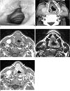
On the direct laryngoscopy, polypoid masses with smooth surfaces were found involving both the true and false vocal folds (Fig. 1A). We suspected the man was suffering from vocal cord carcinoma.
The CT scan of neck showed diffuse thickening involving both the true and false vocal cords without involvement of the anterior commissure and paraglottic space (Fig. 1B). MRI of the neck was performed using a 1.5T imager, and this showed polypoid soft tissue thickening with hypointensity along the true and false vocal cords on the axial T1 (Fig. 1C) and T2-weighted images (Fig. 1D). After infusion of gadolinium contrast agent, this lesion showed homogenous enhancement (Fig. 1E). There was no evidence of destruction of the underlying thyroid cartilage or that the cervical lymph nodes were enlarged.
Biopsy was performed at the true vocal cord lesion. The histopathologic results revealed submucosal amyloid deposition with positive congo red staining. The masses involving the both the false and true vocal cords were resected by performing carbon dioxide laser resection and the patient was discharged without complication.
Discussion
The localized form of amyloidosis is less common (10-20%) than the systemic form. The larynx is the most common site of involvement in the upper aerodigestive tract (123456). The most common site of involvement is the true vocal cords. The false vocal cords (the aryepiglottic folds) and the subglottic region are less commonly involved. The lesions are described as well-defined, submucosal soft-tissue masses, with no or only a slight degree of contrast enhancement. These lesions may also appear as more diffuse intralaryngeal soft tissue infiltration or as a circumferential glottis or subglottic soft tissue thickening, and this is possibly associated with a deeper submucosal component. Areas of localized calcification within the mass are occasionally present (78).
MR imaging demonstrates the specific features of amyloidosis. The amyloid deposit have intermediate T1-weighted signal intensity and low T2-weighted signal intensity are similar to that of skeletal muscle on MR imaging, and this because the amyloid deposits occur as protein fibrils in a parallel, sheet-like configuration that is similar to the organization of skeletal muscle fibers (8).
In our case, the laryngeal amyloidosis presenting as diffuse thickening of the true and false vocal cords was confused with glottic cancer. However, the smooth surface of the lesion seen on the direct laryngoscopy due to the submucosal location and the distinctive hypointensity on the T2-weighted image are helpful findings for making the differential diagnosis. Yet increased signal intensity on T2-weighted images has been also reported (9).
The treatment for localized laryngeal amyloid deposits is surgery using microendoscopic carbon dioxide laser resection (6).
In summary, we report on the imaging of laryngeal amyloidosis that involved the vocal cords. Awareness with the radiologic and clinical characteristics of this disease can help ensure arriving at an accurate diagnosis for patients suffering with laryngeal amyloidosis.
 XML Download