Abstract
Purpose
To evaluate the enhancing solid component within mature ovarian teratomas on pelvic MR examinations.
Materials and Methods
Thirty-two women with surgically proven mature cystic teratomas underwent preoperative pelvic MR examinations. Five cases had an enhancing solid component within mature cystic ovarian teratomas on MR images. The MR images were retrospectively analyzed by two radiologists by consensus, focusing on the enhancing portion of tumor and the tumor itself.
Results
The study subjects include 5 patients (15.6%) with enhancing solid components within the mature ovarian cystic teratomas. The mean tumor size was 9.8 cm and they were all unilateral. The enhancing solid components of the tumors had a variable appearance and were located in the peripheral region. No cases were found to have a transmural extension or direct invasion of the neighboring pelvic organ.
Figures and Tables
Fig. 1
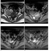
Benign mature cystic teratoma of right ovary in a 40-year-old woman (patient 3 on table 2). Axial T1-weighted (A) and fat-suppression (B) MR images show large lobulated mass containing fat component (arrows). At more caudal level, axial T1-weighted fat-suppression (C) and contrast-enhanced fat-suppression (D) MR images show ovoid enhancing portion (arrows) within the mass.

Fig. 2
Benign mature cystic teratoma of left ovary in a 6-year-old girl (patient 5 on table 2). Axial T1-weighted (A) and fat-suppression (B) MR images show large multiloculated mass containing fat component (arrows). At more cranial level, axial T1-weighted (C) and contrast-enhanced (D) MR images show horseshoe-shaped enhancing portion (arrows) within the mass.

Fig. 3
Benign mature cystic teratoma of left ovary in a 25-year-old woman (patient 4 on table 2). Axial T1-weighted (A) and fat-suppression (B) MR images show large multiloculated ovoid mass containing fat component (arrows). At more caudal level, axial T1-weighted fat-suppression (C) and contrast-enhanced (D) MR images show Y-shaped enhancing portion (arrows) within the mass.

References
1. Kido A, Togashi K, Konishi I, Kataoka ML, Koyama T, Ueda H, et al. Dermoid cysts of the ovary with malignant transformation: MR appearance. AJR Am J Roentgenol. 1999; 172:445–449.
2. Rim SY, Kim SM, Choi HS. Malignant transformation of ovarian mature cystic teratoma. Int J Gynecol Cancer. 2006; 16:140–144.
3. Rha SE, Byun JY, Jung SE, Kim HL, Oh SN, Kim H, et al. Atypical CT and MRI manifestations of mature ovarian cystic teratomas. AJR Am J Roentgenol. 2004; 183:743–750.
4. Sanghera P, El Modir A, Simon J. Malignant transformation within a dermoid cyst: a case report and literature review. Arch Gynecol Obstet. 2006; 274:178–180.
5. Park JY, Kim DY, Kim JH, Kim YM, Kim YT, Nam JH. Malignant transformation of mature cystic teratoma of the ovary: experience at a single institution. Eur J Obstet Gynecol Reprod Biol. 2008; 141:173–178.
6. Ajithkumar TV, Abraham EK, Nair MK. Osteosarcoma arising in a mature cystic teratoma of the ovary. J Exp Clin Cancer Res. 1999; 18:89–91.
7. Park SB, Kim JK, Kim KR, Cho KS. Preoperative diagnosis of mature cystic teratoma with malignant transformation: analysis of imaging findings and clinical and laboratory data. Arch Gynecol Obstet. 2007; 275:25–31.
8. Hackethal A, Brueggmann D, Bohlmann MK, Franke FE, Tinneberg HR, Munstedt K. Squamous-cell carcinoma in mature cystic teratoma of the ovary: systematic review and analysis of published data. Lancet Oncol. 2008; 9:1173–1180.
9. Park SB, Kim JK, Kim KR, Cho KS. Imaging findings of complications and unusual manifestations of ovarian teratomas. Radiographics. 2008; 28:969–983.
10. Mori Y, Nishii H, Takabe K, Shinozaki H, Matsumoto N, Suzuki K, et al. Preoperative diagnosis of malignant transformation arising from mature cystic teratoma of the ovary. Gynecol Oncol. 2003; 90:338–341.
11. Kim JC, Byun JY, Lee YR. MR findings of struma ovarii. J Korean Radiol Soc. 1999; 40:535–541.


 PDF
PDF ePub
ePub Citation
 Print
Print


 XML Download