

 PDF
PDF ePub
ePub Citation
 Print
Print


Abstract
Pulmonary vein varix is a rare vascular abnormality defined as localized pulmonary vein dilatation and is rarely reported in the literature. We report a case of pulmonary vein varix in a 20-year-old man who underwent a screening study for military service. The lesion was shown on CT as a focal dilatation of the pulmonary vein located before draining into the right inferior pulmonary vein and with no connection to the pulmonary artery. This lesion should be included in the differential diagnosis of pulmonary nodules.
Figures and Tables
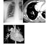
Fig. 1
A. Chest PA shows a 2.3-cm-sized, lobulated-marginated nodule (arrowheads) in right lower lobe, which appears to be connected to adjacent dilated vascular structure.
B. MDCT after administration of contrast media at the level of left ventricle shows localized dilatation of pulmonary vein (arrow) in right lower lobe.
C. 3D reconstruction image shows focally dilated pulmonary vein (arrowheads) in right lower lobe connected to right inferior pulmonary vein. Any connection with pulmonary artery is not seen.
References
1. Fraser RS, Colman NC, Müller NL, Paré PD. Developmental anomalies affecting the pulmonary vessels. Diagnosis of disease of the chest. 4th ed. Philadelphia: W.B. Saunders;1999. 23:p. 645–647.
2. Kumazoe H, Komori M, Ochiai R, Egashira R, Nakazono T, Kudo S. Pulmonary varix mimicking arteriovenous malformation. Clin Imaging. 2008; 32:61–64.
3. Batram C, Strickland B. Pulmonary varices. Br J Radiol. 1971; 44:927–935.
4. Asayama J, Shiguma R, Katsume H, Ijichi H. Pulmonary varix. Angiology. 1984; 35:735–739.
5. Vanherreweghe E, Rigauts H, Bogaerts Y, Meeus L. Pulmonary vein varices: diagnosis with multi-slice helical CT. Eur Radiol. 2000; 10:1315–1317.
 XML Download