

 PDF
PDF ePub
ePub Citation
 Print
Print


Abstract
Purpose
To assess the influence of calcified plaque characteristics on the overestimation of coronary arterial stenosis on a coronary CT angiography (CCTA).
Materials and Methods
The study included 271 coronary arteries with calcified plaques identified by CCTA, and based on 928 coronary arteries from 232 patients who underwent both CCTA and invasive coronary angiography (ICA). Individual coronary arteries were classified into two groups by agreement based on the degree of stenosis from each CCTA and ICA: 1) group A includes patients with concordant CCTA and ICA results and, 2) group B includes patients with an overestimation of CCTA compared to ICA. Parameters including total calcium score, calcium score of an individual coronary artery, calcium burden number of an individual coronary artery, and the density of each calcified plaque (calcium score/number of calcium burden) for each individual coronary artery were compared between the two groups.
Results
Of the 271 coronary arteries, 164 (60.5%) were overestimated on CCTA. The left anterior descending artery (LAD) had a significantly low rate of overestimation (47.1%) compared to the other coronary arteries (p=0.001). No significant differences for total calcium score, calcium score of individual coronary artery, and the density of each calcified plaque from individual coronary arteries between two groups was observed. However, a decreasing tendency for the rate of overestimation on CCTA was observed with an increase in calcium burden of individual coronary arteries (p<0.05).
Conclusion
The evaluation of coronary arteries suggests that the degree of coronary arterial stenosis had a tendency to be overestimated by calcified plaques on CCTA. However, the rate of overestimation for the degree of coronary arterial stenosis by calcified plaques was not significantly influenced by total calcium score, calcium score of individual coronary artery, and density of each calcified plaque.
Figures and Tables
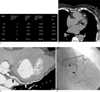 | Fig. 1A 76-year-old woman with chest pain.A. Screenshot of calcium scoring displays number of calcium burden as number of lesion.
B. Precontrast CT image shows multiple calcified plaques in proximal & mid-left circumflex artery.
C. Curved multiplanar reformation of coronary CT angiography (CCTA) image shows significant stenosis (arrows) at proximal & mid-left circumflex artery.
D. Invasive coronary angiography shows corresponding results (arrows) with CCTA.
|
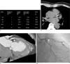 | Fig. 2A 65-year-old man with chest pain.A. Screenshot of calcium scoring displays number of calcium burden as number of lesion.
B. Precontrast CT scan shows dense calcified plaques in proximal left anterior descending artery.
C. Curved multiplanar reformation of coronary CT angiography image shows significant stenosis (arrow) at left anterior descending artery.
D. Invasive coronary angiography shows insignificant (<50%) luminal narrowing (arrow) at proximal left anterior descending artery.
|
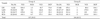
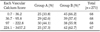
Table 1
The Number of Vessels and Parameters Related with Calcified Plaques of Matched (A) and Overestimated (B) Groups on Coronary CT Angiography (CCTA) according to Location of Calcified Plaques, on Comparison between CCTA and Invasive Coronary Angiography
![]()
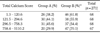
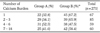
Table 2
Comparison of the Number of Vessels between Matched (A) and Overestimated (B) Groups on CCTA according to Ranked Total Calcium Score as Quartile
![]()
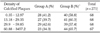
Table 3
Comparison of the Number of Vessels between Matched (A) and Overestimated (B) Group on CCTA according to Ranked Each Vascular Calcium Score as Quartile
![]()
References
1. Stary HC. Evolution and progression of atherosclerotic lesions in coronary arteries of children and young adults. Arteriosclerosis. 1989; 9:1 Suppl. I19–I32.
2. Agostoni P, Biondi-Zoccai GG, De Benedictis ML, Rigattieri S, Turri M, Anselmi M, et al. Radial versus femoral approach for percutaneous coronary diagnostic and interventional procedures. Systematic overview and meta-analysis of randomized trials. J Am Coll Cardiol. 2004; 44:349–356.
3. Budoff MJ, Achenbach S, Blumenthal RS, Carr JJ, Goldin JG, Greenland P, et al. Assessment of coronary artery disease by cardiac computed tomography: a scientific statement from the American Heart Association Committee on Cardiovascular Imaging and Intervention, Council on Cardiovascular Radiology and Intervention, and Committee on Cardiac Imaging, Council on Clinical Cardiology. Circulation. 2006; 114:1761–1791.
4. Hamon M, Biondi-Zoccai GG, Malagutti P, Agostoni P, Morello R, Valgimigli M, et al. Diagnostic performance of multislice spiral computed tomography of coronary arteries as compared with conventional invasive coronary angiography. A meta-analysis. J Am Coll Cardiol. 2006; 48:1896–1910.
5. Stein PD, Beemath A, Kayali F, Skaf E, Sanchez J, Olson RE. Multidetector computed tomography for the diagnosis of coronary artery disease: a systematic review. Am J Med. 2006; 119:203–216.
6. Scheffel H, Alkadhi H, Plass A, Vachenauer R, Desbiolles L, Gaemperli O, et al. Accuracy of dual-source CT coronary angiography: first experience in a high pre-test probability population without heart rate control. Eur Radiol. 2006; 16:2739–2747.
7. Dewey M, Teige F, Schnapauff D, Laule M, Borges AC, Wernecke KD, et al. Noninvasive detection of coronary artery stenoses with multislice computed tomography or magnetic resonance imaging. Ann Intern Med. 2006; 145:407–415.
8. Martuscelli E, Romaqnoli A, D'Eliseo A, Razzini C, Tomassini M, Sperandio M, et al. Accuracy of thin-slice computed tomography in the detection of coronary stenoses. Eur Heart J. 2004; 25:1043–1048.
9. Hoffmann MH, Shi H, Schmitz BL, Schmid FT, Lieberknecht M, Schulze R, et al. Noninvasive coronary angiography with multislice computed tomography. JAMA. 2005; 293:2471–2478.
10. Hoffmann U, Moselewski F, Cury RC, Ferencik M, Jang IK, Diaz LJ, et al. Predictive value of 16-slice multidetector spiral computed tomography to detect significant obstructive coronary artery disease in patients at high risk for coronary artery disease: patient-versus segment-based analysis. Circulation. 2004; 110:2638–2643.
11. Hoffmann U, Moselewski F, Nieman K, Jang IK, Ferencik M, Rahman AM, et al. Noninvasive assessment of plaque morphology and composition in culprit and stable lesions in acute coronary syndrome and stable lesions in stable angina by multidetector computed tomography. J Am Coll Cardiol. 2006; 47:1655–1662.
12. Kroft LJ, De Roos A, Geleij J. Artifacts in ECG-Synchronized MDCT Coronary Angiography. AJR Am J Roentgenol. 2007; 189:581–591.
13. Leschka S, Alkadhi H, Plass A, Desbiolles L, Grünenfelder J, Marincek B, et al. Accuracy of MSCT coronary angiography with 64-slice technology: first experience. Eur Heart J. 2005; 26:1482–1487.
14. Raff GL, Gallagher MJ, O'Neill WW, Goldstein JA. Diagnostic accuracy of noninvasive coronary angiography using 64-slice spiral computed tomography. J Am Coll Cardiol. 2005; 46:552–557.
15. Ong TK, Chin SP, Liew CK, Chan WL, Seyfarth MT, Liew HB, et al. Accuracy of 64-row multidetector computed tomography in detecting coronary artery disease in 134 symptomatic patients: Influence of calcification. Am Heart J. 2006; 151:1323.e1–1323.e6.
16. Kuettner A, Kopp AF, Schroeder S, Rieger T, Brunn J, Meisner C, et al. Diagnostic accuracy of multidetector computed tomography coronary angiography in patients with angiographically proven coronary artery disease. J Am Coll Cardiol. 2004; 43:831–839.
17. Sun Z, Lin CH, Davidson R, Dong C, Liao Y. Diagnostic value of 64-slice CT angiography in coronary artery disease: a systematic review. Eur J Radiol. 2008; 67:78–84.
18. Geleijns J, Kroft LJM, Bax JJ, Lamb HJ, de Roos A. Techniques for cardiovascular computed tomography. In : Higgins CB, de Roos A, editors. MRI and CT of the cardiovascular system. 2nd ed. Philadelphia, PA: Lippincott Williams & Wilkins;2005. p. 37–52.
19. Barrett JF, Keat N. Artifacts in CT: recognition and avoidance. Radiographics. 2004; 24:1679–1691.
20. Sarwar A, Rieber J, Mooyaart EA, Seneviratne SK, Houser SL, Bamberg F, et al. Calcified plaque: measurement of area at thin-section flat-panel CT and 64-section multidetector CT and comparison with histopathologic findings. Radiology. 2008; 249:301–306.
 XML Download