

 PDF
PDF ePub
ePub Citation
 Print
Print


Sinusitis is a common condition; however, serious complications associated with sinusitis, such as osteomyelitis, subperiosteal scalp abscess, orbital cellulitis, and intracranial extension have become quite rare with the advent of antibiotics (1). Occasionally, serious complications may occur with mild symptomatic presentation. In such cases, prompt medical and surgical therapy is required to minimize potentially serious sequelae. Pott's puffy tumor is an unusual but important complication of frontal sinusitis and may occur as a result of the spread of sinusitis to the frontal bone. Recognition of this entity is important, because of the risk of intracranial extension and its attendant complications (2). We present a patient who had clinical signs of diplopia, blurred vision, and mild tenderness on glabellar area. A CT examination showed osteomyelitis of the frontal bone as well as frontal sinusitis.
Case Report
A 64-year-old male patient presented with complaints of diplopia, blurred vision, and mild tenderness on the glabellar area over the previous 25-days. The patient had no history of trauma, however did have a general history of hypertension and a 5-year history of stroke. Upon physical examination, swelling and edema of the right periorbital was observed, which caused diplopia.
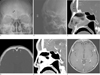
On the PNS series, a blurred margin of the right frontal sinus wall (Fig. 1A) and soft tissue swelling on prefrontal area were seen (Fig. 1B). A CT scan of the orbits and sinuses revealed full opacification of the frontal sinus, erosive thinning of the anterior wall of the frontal sinus (Fig. 1C), sclerotic bone marrow of the frontal skull (Figs. 1C, D) as well as soft tissue swelling and enhancement over the forehead (Fig. 1E).
The patient was diagnosed with frontal sinusitis, forehead, periorbital cellulitis, and osteomyelitis of the frontal calvarium. The otolaryngology service was performed on the trephination of the frontal sinus and a large amount of pus in the frontal sinus was seen with erosion of the frontal bone. The microbiology broth isolated streptococcus viridans.
On contrast enhanced brain magnetic resonance imaging (MRI) performed four months later, enhancement of the frontal bone marrow was seen, however no intracranial abnormal findings were present (Fig. 1F).
Discussion
Pott's puffy tumor was first described as "a puffy, circumscribed, indolent tumor of the scalp and a spontaneous separation of the pericranium from the skull under such a tumor" by Percivall Pott, in London in 1760 (34). The disorder is an eponym for frontal osteomyelitis and is associated with a subperiosteal abscess. With the advent of antibiotics, this disorder is considered to be extremely rare (4).
Sir Percival Pott originally described this condition as a complication of trauma; however, it is more commonly observed as a complication of frontal sinusitis (5). More rare causes of Pott's puffy tumor include insect bites, a craniotomy, or hair transplantation (5). Pott's puffy tumor can occur as a result of the spread of sinusitis to the frontal bone, along with the development of osteomyelitis in the frontal bone and the extension of purulent material (2). Trauma to the prefrontal region of the skull and surrounding soft tissues can also be a cause of the disease (6). When forehead swelling after trauma is associated with fever and sinusitis, clinical suspicion should be raised for the occurrence of Pott's puffy tumor.
Pott's puffy tumor presents with symptoms including frontal scalp swelling, fever, headache, frontal sinus tenderness, and photophobia (1). The frontal scalp swelling may be less tender or less erythematous than expected due to the depth of the infection (1). Headaches are often relieved as the sinus drains through the frontal bone. Occasionally, the patient's symptoms are diminished as the forehead enlarges (6).
Pott's puffy tumor can be associated with intracranial complications such as subdural empyema, an epidural abscess, and cortical vein thrombosis with or without direct erosion of the frontal bone. Because the mucosal venous drainage of the frontal sinus occurs through the diploic veins, which communicate with the dural venous plexus, septic thrombi can potentially evolve from foci within the frontal sinus and propagate through this venous system (25). Also, because the frontal sinus shares a thin posterior plate with the frontal cranial fossa, when the frontal sinusitis spreads to the brain; it most commonly results in an infection of the frontal lobe (7). An orbital subperiosteal abscess, which is a known complication of adjacent sinusitis, can be associated with frontal sinusitis and has been shown to place the patient with orbital cellulitis at a greater risk of intracranial involvement (7).
In previously reported cases, the cultured etiologic organisms included microaerophilic streptococci such as alpha-hemolytic streptococcus, staphylococcus, peptostreptococcus, bacteroides species, and other anaerobes. Because of the relatively anaerobic conditions in the frontal sinus caused by compromised ostial patency, microaerophilic organisms may be more common in the frontal sinusitis (25).
Pott's puffy tumor cannot be excluded without proper imaging studies. A contrast-enhanced head and sinus CT scan is the diagnostic modality of choice (1). Bone scanning will detect osteomyelitis.
Pott's puffy tumor can be differentiated from a mucocele and a malignancy. On a CT scan, a mucocele is expansile and causes an erosive change of the surrounding bony structure; however, no osteomyelitis or soft tissue inflammatory changes are seen. In malignant paranasal sinus lesions, well enhancing soft tissue lesions, bizarre shaped surrounding bony structure destruction, and soft tissue infiltration are seen.
Antibiotic therapy alone is rarely adequate for the treatment of Pott's puffy tumor. The definitive treatment consists of a combination of surgery, including debridement and removal of the sequestrum, as well as treatment with antibiotics to prevent further suppurative complications (14).
Pott's puffy tumor is a complicated infection that requires hospital admission and aggressive therapy. Patients with Pott's puffy tumor generally don't appear acutely ill and have a subtle symptomatic presentation. Therefore, proper imaging diagnosis, such as a contrast-enhanced CT scan, is critical to prevent aggravation of the complications associated with Pott's puffy tumor (1).
This case report describes simple X-ray, CT, and MR imaging findings of Pott's puffy tumor.
 XML Download