

 PDF
PDF ePub
ePub Citation
 Print
Print


INTRODUCTION
Although the pathophysiology of type 2 diabetes mellitus (T2DM) is currently explained by a total of eight pathophysiology, main two pathophysiology of T2DM is insulin secretion defect and insulin resistance [1]. In Western T2DM patients, insulin resistance occurs 10 to 15 years prior to the development of T2DM, and compensatory hyperinsulinemia occurs during the prediabetes stages [2]. However, only about half of Korean T2DM patients are obese. According to euglycemic clamp studies in Korea, a considerable portion of Korean T2DM patients did not have insulin resistance [3]. In addition, the degree of insulin resistance in Korean T2DM patients who have insulin resistance is not as severe as Western T2DM patients [4]. Hence, it is believed that the pathophysiology of Korean T2DM is different from Western T2DM [5]. That being said, the prevalence of obesity and insulin resistance has increased in Korean T2DM during the past three decades. Some groups assert that the main pathophysiology of Korean T2DM are changed and simulate to pathophysiology of Western T2DM [6].
The purpose of this article is to find the main cause of Korean T2DM, either insulin resistance or insulin secretion, and compare the pathophysiology of Korean T2DM with that of Western T2DM. Further, we would like to investigate whether the main cause of Korean T2DM has changed since 1980. We refer to our study results to provide helpful information for establishing treatment strategies in Korean T2DM.
EPIDEMIOLOGY
Prevalence of T2DM in Korea
There has been an abrupt increase in the prevalence of T2DM since the 1960s; westernization and industrialization has also occurred in South Korea during this time period. The prevalence of T2DM was estimated as 0.5% in the 1960s [5]. In a study conducted in 1971, the prevalence was 1.5% (n=15,853) [7], and the prevalence during 1980 to 1983 was 3.5% (n=18,201) according to another study conducted in a health care center [8]. In Yoncheon, a cohort study conducted in 1993 determined that the prevalence was 7.2% (n=2,520) [9], and in a Jungup cohort study conducted in 1998, the prevalence was 8.5% (n=774) [10]. In terms of large-scale prevalence, a prospective community-based cohort study conducted in 2001 to 2002 showed that the prevalence was 12.6% (n=10,038) [11]. The prevalence was 9.9% in Korean National Health and Nutrition Examination Survey (KHANES) 2007 to 2009 [12]. In 2013, the prevalence was 8.0% based on health care insurance data (n=13,512) [13]. Hence, the prevalence of T2DM has increased until the late 1990s and plateaued during the early 2000s at around 10%.
Change of prevalence of obesity in Korean T2DM patients
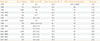
Some articles described that the prevalence of non-obese T2DM in 1980s Korea was around 80% [514], but there was only one article that reported the prevalence of T2DM was closer to 80% (76.7%) [14]. However, in that article, the body mass index (BMI) criteria of obesity in males was 27 kg/m2. So, the prevalence of non-obese T2DM was overestimated. The prevalence of obesity in Korean T2DM is summarized in Table 1 [6141516171819202122232425]. If these two studies where the BMI criteria were not 25 kg/m2 are excluded, it can be seen that the prevalence of non-obese T2DM was actually 60% to 69% in the 1980s and decreased to 50% to 55% in the 2000s. BMI was changed from about 23 kg/m2 in the 1980s to approximately 25 kg/m2 in the 2000s.
PATHOPHYSIOLOGY OF KOREAN T2DM
Heterogeneity in the pathogenesis of Korean T2DM
There was only one report that evaluated insulin secretion function in Korean T2DM in the 1970s and 1980s [2627]. In that report, insulin secretion was lower in normal Korean subjects compared to normal subjects from the West. The first study regarding insulin resistance based on dynamic methods in Korean T2DM was conducted in 1994 [28]. Kim et al. [28] evaluated insulin sensitivity in 22 subjects (mean BMI 23.4 kg/m2) using the euglycemic hyperinsulinemic clamp method. Eight subjects among 22 subjects did not have insulin resistance. In another study conducted in 1998 using the euglycemic hyperinsulinemic clamp method, 22 among 38 T2DM patients were insulin resistant. Fifty percentage of non-obese T2DM and 30% of obese T2DM did not have insulin resistance [29]. These findings suggest that there is heterogeneity in the pathogenesis of Korean T2DM patients.
Role of insulin resistance in Korean T2DM
It is well known that one of the main causes of insulin resistance is obesity. Subjects with T2DM have a higher BMI than normal subjects according to numerous studies. In the KHANES between 2009 and 2013, the prevalence of obesity (BMI ≥25 kg/m2) was 30.0% to 31.8% in the adult general population. The prevalence of obesity in T2DM during this same period was 50% (mean BMI 25.0 to 25.64 kg/m2) [13]. It is suggested that obesity, an indirect marker of insulin resistance, is associated with T2DM.
The prevalence of T2DM with insulin resistance has increased with increasing BMI during the past three decades. One study compared two cross-sectional periods (1997 to 1999 vs. 2007 to 2011) from the same single center. According to a study from the 2000s, homeostatic model assessment of insulin resistance (HOMA-IR), a marker for insulin resistance, was increased compared to that of the 1990s [22]. The Quicki index (quantitative insulin-sensitivity check index) and the Matsuda index, both markers of insulin sensitivity, were decreased. However, insulin secretion parameters (insulinogenic index [IGI], oral disposition index) did not change in T2DM. This means that the contribution of insulin resistance in the pathogenesis of Korean T2DM patients has increased during past decade.
There were two cross-sectional studies that reported that the main pathophysiology in Korean T2DM was insulin resistance. Kim et al. [24] conducted a short insulin tolerance test in T2DM patients in 2004 to 2005. The criteria for insulin resistance was Kitt (≤2.5) that was verified through a normoglycemic-hyperinsulinemic clamp study in Korean T2DM. The prevalence of insulin resistance was 70.6%. Son et al. [6] reported that the prevalence of insulin resistance (HOMA-IR >2.5) was 59.5% according to a SURPRISE study (nation-wide, cross-sectional primary care unit-based study, n=1,314) conducted in 2009 to 2010. The number of patients with insulin resistance was larger than patients with insulin secretion defect (59.5% vs. 22.0%, respectively). Considering these results, it is suggested that insulin resistance contributes to the development of T2DM in many Korean populations.
Role of insulin secretion defect in Korean T2DM
Yoon [30] conducted an oral glucose tolerance test (OGTT) in 450 subjects that were comprised of normal glucose tolerance (NGT) patients and patients with diagnosis of prediabetes and diabetes in 1995. In both the obese and non-obese groups, even within normal fasting glucose, 30 minutes insulin of OGTT declined as fasting glucose rise. Rhee et al. [31] reported similar results in 2010 (n=873). The disposition index calculated from OGTT declined as fasting glucose rise even within normal fasting glucose. Also, in other methods that use euglycemic-hyperinsulinemic clamps, both the obese and non-obese T2DM showed an insulin secretion defect (n=38, conducted in 1998) [29]. These results suggest that insulin secretion defect occurs in early stages, even with NGT, not only in the non-obese group but also in the obese group. Compensatory β-cell response was not observed in obese groups.
One study used a frequent sampled intravenous glucose tolerance test (FSIVGTT) for NGT and pre-diabetes, conducted in 1998 (n=60) [32]. Studying prediabetes is valuable because it can indicate early abnormalities in the development of T2DM. FSIVGTT is more accurate method than OGTT for the evaluation of insulin resistance and secretion. There were no differences in the insulin sensitivity index (SI) between the NGT, non-obese impaired glucose tolerance (IGT), and obese IGT groups (0.70±0.25, 0.61±0.39, and 0.39±0.39 ×10⁻⁴/min/pmol/L, respectively). The acute insulin response to glucose (AIRg), an index of insulin secretion, was decreased in both the non-obese and obese IGT compared to NGT (164±155, 200±195, and 367±230 pmol/L, respectively). In 2003, that was a similar study using FSIVGTT with 40 subjects comprised of NGT, IGT, and T2DM groups [33]; these results were similar to a previous study. There was no difference in the insulin SI between the three groups. The insulin secretion index (AIRg) was decreased in the IGT group when compared with NGT. AIRg was much lower in the T2DM group than the IGT group. This means that regardless of obesity, the initial pathophysiologic abnormality in IGT was insulin secretion defect in Korea.
Kim et al. [23] conducted an OGTT for 370 subjects that were comprised of NGT, prediabetes and T2DM patients during 1997 to 2000. Between combined glucose intolerance (CGI) and T2DM, there was no difference in HOMA-IR. In the obese group, there was no difference in HOMA-IR between NGT, impaired fasting glucose (IFG), IGT, CGI, and T2DM. As the glucose tolerance state progresses from NGT to T2DM (NGT, IFG, IGT, CGI, and T2DM), insulin secretion (IGI) steadily decreased, regardless of obesity. This means that the initial abnormality leading to diabetes in both the obese and non-obese group was insulin secretion defect in Korea T2DM.
Recently new longitudinal study for incident T2DM in Korean non-diabetic subjects were published [34]. In that study, index for insulin resistance (HOMA-IR) did not predict incident T2DM. But, insulin secretion indexes (insulinogenic index and C-peptidogenic index) could predict incident T2DM.
Criticism of studies that report insulin resistance is more important
Kim et al. [24] and Son et al. [6] used fasting C-peptide as a marker of insulin secretion. Patients with fasting serum C-peptide concentrations <1.1 ng/mL (0.37 nmol/L), 1.1 to 1.7 ng/mL (0.37 to 0.56 nmol/L), or more than 1.7 ng/mL (0.57 nmol/L) were classified as having a severe secretory defect, moderate secretory defect, or mild to no secretory defect, respectively. However, there are some limitations to these criteria. First, there is only one reference study that verified these criteria as a secretory function; in addition, the main focus of this study was not about fasting C-peptide and insulin secretion, but instead was the short insulin tolerance test. Second, C-peptide more than 1.7 ng/mL cannot discriminate against the mild secretory defect group and no secretory defect group. In the OGTT study that Kim et al. [10] performed, baseline C-peptide was not different amongst the NGT, prediabetes and T2DM groups. When we analyzed our data, among the 89 prediabetes subjects who had a fasting C-peptide higher than 1.7 ng/mL, 40.5% had insulin secretion defect (IGI <0.4). Generally, fasting insulin solely is not used as an insulin secretion index. The same principle can be applied to fasting C-peptide, and fasting C-peptide is a very rough index for insulin secretion.
Son et al. [6] reported that 20.2% of patients exhibited a moderate insulin secretion defect and 59.5% of subjects had insulin resistance. However, it cannot be concluded that main pathogenesis of T2DM has shifted from insulin deficiency to insulin resistance in the Korean population. Among 79.8% of subjects who had a fasting C-peptide higher than 1.7 ng/mL, we cannot know which percentage of subjects have had an insulin secretory defect. Additionally, HOMA-IR can be overestimated in the T2DM group. Thus, it's better to use HOMA-2 IR as an insulin resistance marker in the diabetes group.
Both insulin resistance and secretion defect have a role for the development of T2DM in Korea
There have been very important limitations in discussing the role of insulin resistance and insulin secretion in the development of Korean T2DM. First of all, there have been only cross-sectional studies. In 2001 to 2002, the Korean government funded a large-scale, community-based prospective cohort study (the Korean Genome and Epidemiology Study) that observed the same people longitudinally. This study followed up on 4,106 participants with NGT using OGTTs every 2 years for 10 years [35]. They used the Matsuda index as a marker of insulin resistance and the 60 minutes IGI (IGI60) as a marker of insulin secretion. They classified the study participants into three groups (the NGT group, proceed to prediabetes group, and proceed to diabetes group) after 10 years of follow-up. In all three groups, insulin resistance was increased. Baseline HOMA-IR was not different between the progressor to diabetes and the non-progressor. However, the Matsuda index was lower in the progressor group. The NGT groups showed compensatory increased insulin secretion, but the progress to diabetes group showed no change in the insulin secretion index during the follow-up period. In the progress to prediabetes group and the progress to diabetes group, the oral disposition declined during follow-up period. Baseline IGI60 was lower in the progressor group compared to the non-progressor group. These results suggest that both insulin resistance and insulin secretion defect contribute to the development of T2DM in Korean people.
It is important to note that Korean T2DM did not show compensatory insulin secretion under the conditions of insulin resistance, contrary to Western T2DM, who had a compensatory hyperinsulinemia stage before the development of T2DM. It may be summarized that insulin resistance is a necessary condition. However, insulin secretion defect is a sufficient condition for the development of Korean T2DM, in mathematical terms. Insulin resistance provides an opportunity for β-cells to develop β-cell dysfunction. If β-cells compensate, they do not develop T2DM. If not, they will develop T2DM in the Korean population.
These findings were consistent with previous findings in two cross-sectional studies. One study previously mentioned compared two cross-sectional periods in the same single center (1990s vs. 2000s) [22]. They compared 504 subjects (mean age, 50.2 years, recruited in 1997 to 1999) and 578 subjects (mean age 48.5 years, recruited in in 1997 to 2011). In the 2000s, HOMA-IR, a marker of insulin resistance was increased compared to the 1990s (1.45±0.11 vs. 1.75±1.75, P=0.002 in the NGT group; 1.91±0.11 vs. 2.29±0.10, P=0.013 in the pre-diabetes group; 2.63±0.10 vs. 2.95±0.11, P=0.017 in the diabetes group). The Quicki index and the Matsuda index, markers of insulin sensitivity, were both decreased. However, other insulin secretion parameters (IGI 23.31±1.59 vs. 27.32±1.68, P=0.061), oral disposition index (0.44±0.05 vs. 0.57±0.72, P=0.103) did not changed in diabetes group.
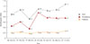
In another cross-sectional study, Rhee et al. [31] performed OGTT with 873 subjects who were suspected of abnormal glucose tolerance. Although, in prediabetes, insulin resistance was increased (HOMA-IR, 1.90±0.20 in NGT vs. 2.33±0.11 in prediabetes, P<0.001; Matsuda index, 0.73±0.01 in NGT vs. 0.63±0.11 in prediabetes, P<0.001) compared to NGT, insulin secretion was decreased (IGI, 1.25±0.07 in NGT vs. 0.85±0.004 in prediabetes, P<0.001; HOMA-β, 120.2±9.9 in NGT vs. 82.1±5.6 in prediabetes, P<0.001). It means that normal physiology response (compensatory increased insulin secretion) to insulin resistance were not observed in Korean T2DM. The same finding are seen in Fig. 1 [222325313637], which is a graph of the IGI in Korean T2DM using retrospectively collected data from several cross-sectional studies over the span of two decades. Increased trends were observed in the IGI of NGT and prediabetes groups. However, it seems that there was no definite change in the diabetes group.
CONCLUSIONS
It is unreasonable to focus on only one of insulin resistance and insulin secretion. Most T2DM patients have both insulin resistance and insulin secretion defect. In many Korean T2DM patients, insulin resistance contributes to T2DM. If insulin resistance develops in people that are genetically susceptible to having low β-cell function, and if β-cells do not compensate for insulin resistance appropriately, the patient will develop T2DM. Insulin resistance makes people more prone to develop T2DM, and Koreans become T2DM easily with a relatively low BMI condition. The increased prevalence of insulin resistance may contribute to the increased prevalence of T2DM during the past three decades in Korea. Current evidence suggests that insulin resistance is a contributing factor and insulin secretion defect is a determinant for the development of T2DM in Koreans.
 XML Download