Journal List > Endocrinol Metab > v.31(2) > 1086254

Wiersinga: Clinical Relevance of Environmental Factors in the Pathogenesis of Autoimmune Thyroid Disease

Abstract

Genetic factors contribute for about 70% to 80% and environmental factors for about 20% to 30% to the pathogenesis of autoimmune thyroid disease (AITD). Relatives of AITD patients carry a risk to contract AITD themselves. The 5-year risk can be quantified by the so-called Thyroid Events Amsterdam-score, based on serum thyroid-stimulating hormone, thyroid peroxidase (TPO)-antibodies and family history. Subjects at risk may ask what they can do to prevent development of AITD. This review summarizes what is known about modulation of exposure to environmental factors in terms of AITD prevention. To stop smoking decreases the risk on Graves disease but increases the risk on Hashimoto disease. Moderate alcohol intake provides some protection against both Graves and Hashimoto disease. Low selenium intake is associated with a higher prevalence of thyroid autoimmunity, but evidence that selenium supplementation may lower TPO antibodies and prevent subclinical hypothyroidism remains inconclusive. Low serum vitamin D levels are associated with a higher prevalence of TPO antibodies, but intervention studies with extra vitamin D have not been done yet. Stress may provoke Graves hyperthyroidism but not Hashimoto thyroiditis. Estrogen use have been linked to a lower prevalence of Graves disease. The postpartum period is associated with an increased risk of AITD. Taking together, preventive interventions to diminish the risk of AITD are few, not always feasible, and probably of limited efficacy.

INTRODUCTION

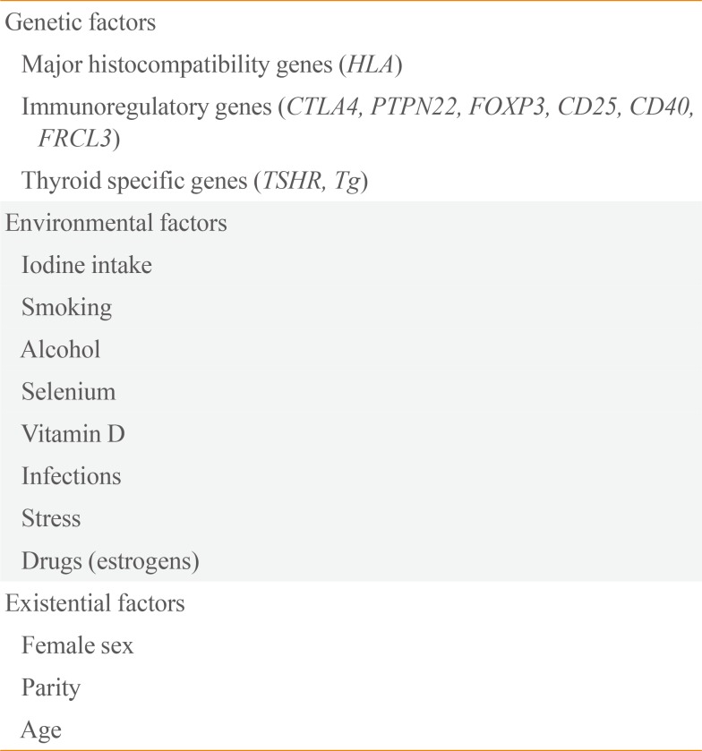
Autoimmune thyroid diseases (AITD) like Graves disease and Hashimoto disease are complex diseases in which autoimmunity against thyroid autoantigens develop against a certain genetic background, provoked by exposure to environmental factors. Weetman [1] has used Swiss cheese as a model to explain the immunopathogenesis of AITD. Swiss cheese has characteristic big holes. Assuming each hole represents a genetic, environmental, or existential factor, the disease may manifest itself when a number of subsequent holes are aligned in a way that an arrow may pass them all in one shot. Table 1 lists these various factors. Thus the autoimmune reaction is facilitated by polymorphisms in major histocompatibility genes (human leukocyte antigen [HLA]), immunoregulatory genes, and thyroid specific genes, probably provoked by various environmental factors and enhanced by existential factors like female gender, parity, and age. Twin studies have indicated that the relative contribution of genetic factors to the development of Graves' hyperthyroidism is about 79% [2]. Likewise, genetic factors would contribute for about 73% to the development of thyroid peroxidase (TPO) antibodies and/or Tg antibodies [34]. The implication is that the proportion of environmental factors in the immmunopathogenesis of AITD is limited, in the order of 20% to 30%. The number of known susceptibility loci increases every year, but taken together accounts for less than 20% of the heritability of AITD [15]. Presumably many more unidentified susceptibility loci are involved.
AITD frequently runs in families, and having a family member with AITD increases the risk you yourself will get AITD. The risk for siblings of AITD patients can be quantified by the sibling risk ratio λs, defined as the risk for developing AITD in a sibling of an AITD patient divided by the frequency of AITD in the general population. Λs is 16.9 for AITD, 11.6 for Graves' hyperthyroidism, and 28.0 for Hashimoto's hypothyroidism [6]. The λs is a rather rough risk estimate for individual subjects. I have been frequently asked by patients in whom I had just diagnosed overt Graves or Hashimoto disease: "Doctor, I know my thyroid disease runs frequently in families and occurs especially in women. I am concerned about my daughter who is now in her twenties, and planning to become pregnant some years from now. How high is the chance she will also get thyroid disease, and can anything be done to prevent that happening?" Prevention in this context would mean modulation of exposure to particular environmental factors, as changing the genetic make-up of an individual albeit theoretically possible is not that easy. Preventive interventions are likely to be more effective in persons who already carry a somewhat higher risk. Consequently in this review article we will first evaluate the predictability of developing AITD, and thereafter review the role of various environmental factors in the development of AITD. Lastly, a synopsis will be provided what an individual at risk to develop AITD, can do to diminish that risk.

PREDICTION OF AUTOIMMUNE THYROID DISEASE

The Amsterdam AITD cohort is a 5-year follow-up study in a population at risk for AITD, namely in healthy women with one or more 1st or 2nd degree relatives with proven AITD [7]. The study aim was to design a predictive score for the development of overt autoimmune hypo- or hyperthyroidism, and to learn more about the pathogenesis of AITD especially on the role of environmental factors in the early stages of the disease. At study entrance women should be without known thyroid disease and without overt hyper- or hypothyroidism. There were 790 participants with a mean age of 36±12 years and median thyroid-stimulating hormone (TSH) was 1.7 mU/L. Subclinical hyperthyroidism was detected in 13 and subclinical hypothyroidism in 19 participants at baseline. TPO-Ab ≥100 kU/L were present in 27%, Tg-Ab ≥100 kU/L in 8%, a family history of Graves disease in 76% and of Hashimoto disease in 49%. Participants were seen annually for blood sampling and filling in questionnaires. After 5-year follow-up, there had been 38 events of overt hypothyroidism (34×Hashimoto's thyroiditis, 4×postpartum thyroiditis) and 13 events of overt hyperthyroidism (11×Graves' hyperthyroidism, 1×postpartum thyroiditis, and 1×silent thyroiditis). The mean annual event rate was 1.5%, and the 5-year cumulative event rate was 7.5%. The incidence rate per 1,000 women years in the Amsterdam AITD cohort was 9.6 for overt hypothyroidism and 3.3 for overt hyperthyroidism; these figures are about three times higher than observed in population-based studies in the United Kingdom (UK): 3.5 and 4.98 for overt hypothyroidism and 0.8 and 0.77 for overt hyperthyroidism in the Whickham and Tayside regions of the UK, respectively [89].
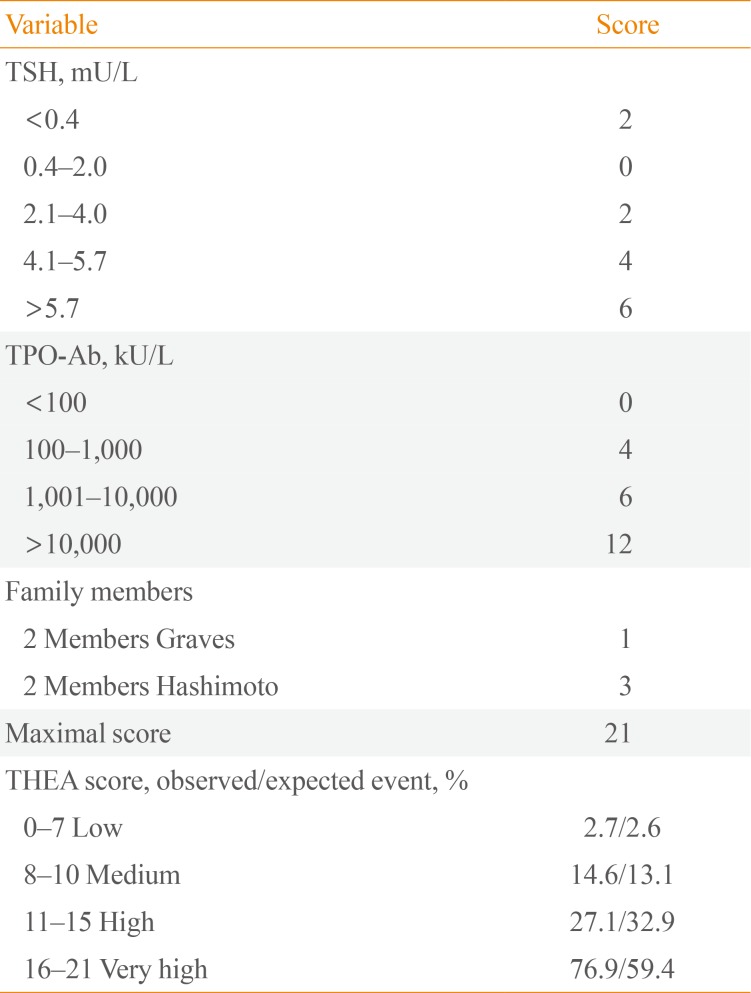
Independent risk factors for the occurrence of overt hyper- or hypothyroidism in the Amsterdam AITD cohort were baseline TSH, baseline TPO-Ab, and family background of AITD. A predictive score for such events was obtained from a logistic regression model by putting weights to individual risk factors proportional to their relative risk. This so-called Thyroid Events Amsterdam (THEA) score is listed in Table 2. Low scores are associated with a low incidence of overt hyper- or hypothyroidism in the next 5 years, and vice versa [7].
Getting back to the patient who was concerned about her 23-year old daughter who wanted to become pregnant in the near future, her mother had Graves' hyperthyroidism and her grandmother Hashimoto's hypothyroidism. Thus the daughter scores zero for "family members." Blood sampling was done in the daughter, revealing TSH 2.5 mU/L and TPO-Ab 500 kU/L; the resulting THEA score is 6, meaning a low chance of about 2.7% for developing overt thyroid dysfunction in the next 5 years. Would her TSH have been 6.0 mU/L and her TPO-Ab 1,100 kU/L, then her THEA score of 12 would indicate a much higher risk of about 30%. The next question obviously is whether she can do anything by herself in order to decrease that risk.

ENVIRONMENTAL FACTORS IN THE PATHOGENESIS OF AUTOIMMUNE THYROID DISEASE

Iodine intake

It is well known that at a population level thyroid antibodies and autoimmune hypothyroidism are more common in iodine-replete areas than in iodine-deficient areas. The most recent evidence supporting this statement comes from Denmark. The prevalence of TPO-Ab before and after mandatory iodization of salt was 14.3% and 23.8%, respectively [10], and the incidence of overt hypothyroidism increased from 38.3/100.000 per year at baseline to 47.2/100.000 per year 5 to 7 years after iodine fortification of salt [11]. Voluntary iodine prophylaxis in a small Italian community also increased prevalence of TPO-Ab (12.6% vs. 19.5%) and hypothyroidism (2.8% vs. 5.0%) 15 years later [12]. At an individual level, one should follow the World Health Organization recommendation of a daily iodine intake of about 150 µg.

Smoking

It has been known for a long time that smoking is a risk factor for Graves disease [13]. Current smoking increases the risk of Graves' hyperthyroidism about two-fold and of Graves' orbitopathy about three-fold. The effect is dose-dependent, more pronounced in women than in men, and disappears a few years after cessation of smoking. Less well known is the more recent finding that smoking to a certain extent protects against hypothyroidism. Current smokers have a lower prevalence of TPO-Ab as compared to non-smokers, both in the Amsterdam AITD cohort at baseline (odds ratio [OR], 0.69; 95% confidence interval [CI], 0.48 to 0.99) and in the third National Health and Nutrition Examination Survey (OR, 0.57; 95% CI, 0.48 to 0.67) [1415]. In the population-based HUNT study in Norway current smokers as compared to never smokers have also a lower prevalence of subclinical hypothyroidism (OR, 0.54; 95% CI, 0.45 to 0.66) and of overt hypothyroidism (OR, 0.60; 95% CI, 0.38 to 0.95) [16]. The effect is dose-dependent, and disappears a few years after cessation of smoking. In the longitudinal Amsterdam AITD cohort de novo occurrence of TPO-Ab and/or Tg-Ab occurred in 20% of the initially antibody-negative participants during the 5-year follow-up [17].
Participants who stopped smoking in this period increased their risk to develop de novo TPO-Ab and/or Tg-Ab. In the prospective DanThyr study, 140 patients diagnosed with autoimmune hypothyroidism had more often quitted smoking in the last 2 years before diagnosis than 560 matched controls (16.4% vs. 3.4%) [18]. The increased risk of autoimmune hypothyroidism upon quitting was transient: OR's <1 year after stopping smoking, 1 to 2 years after cessation of smoking and 3 to 10 years after quitting were 7.36 (95% CI, 2.27 to 23.90), 6.34 (95% CI, 2.59 to 15.3), and 0.75 (95% CI, 0.30 to 1.87), respectively.

Alcohol

A nested case-control study in the Amsterdam AITD cohort did not find a relationship between alcohol consumption and de novo development of TPO-Ab, but participants who developed overt autoimmune hypothyroidism consumed less alcohol than those who remained euthyroid [19]. A population-based case-control study in Denmark likewise observed that moderate alcohol consumption reduces the risk of overt autoimmune hypothyroidism: OR's were 1.98 (95% CI, 1.21 to 3.33) for 0 alcoholic units/week, 1.00 for 1 to 10 units/week (reference), 0.41 (95% CI, 0.20 to 0.83) for 11 to 20 units/week, and 0.90 (95% CI, 0.41 to 2.00) for ≥21 units/week [20]. Most remarkable, the same group of Danish investigators showed that moderate alcohol consumption also reduces the risk of Graves' hyperthyroidism: OR's were 1.73 (95% CI, 1.17 to 2.56) for 0 units/week, 1.00 for 1 to 2 units/week (reference), 0.56 (95% CI, 0.39 to 0.79) for 3 to 10 units/week, 0.37 (95% CI, 0.21 to 0.65) for 11 to 20 units/week, and 0.22 (95% CI, 0.08 to 0.60) for ≥21 units/week [21]. The observed associations were independent of the type of alcohol (wine or beer did not matter), gender, iodine intake, or smoking behaviour. As there exists a clear association between smoking and alcohol intake, smoking could have been an important confounder in these studies, but that was not observed. The observed association between alcohol intake and AITD likely indicates a cause-and-effect relationship in view of the strength and consistency of the associations and the presence of a dose-response effect. A protective effect of alcohol has also been recorded for other autoimmune diseases like rheumatoid arthritis and type 1 diabetes. The mechanism of action of these protective effects of alcohol remain poorly understood.

Selenium

Glutathione peroxidases and thioredoxin reductases are selenoproteins involved in regulation of the redox state and protection from oxidative damage. The thyroid gland contains more selenium per gram tissue than any other organ. The enzyme GP×3 protects thyrocytes from oxidative stress generated by the action of H2O2. Low selenium levels have been associated with poor immune function. Thus it has been hypothesized that mild nutritional selenium deficiency may promote thyroid autoimmunity. Conversely, selenium supplementation might have a beneficial effect on thyroid autoimmunity, and that has been tested in up to now eight randomized placebo-controlled clinical trials (Table 3). Recruited were patients with Hashimoto's thyroiditis and TPO antibodies. Baseline TSH was either normal or slightly elevated, and exogenous levothyroxine was used by participants in some of the trials. Primary outcome was change in TPO-Ab, which makes sense. For, in the natural history of AITD, TPO-Ab are detectable in serum for a long time (actually for years) with a gradual increase in concentration before serum TSH becomes slightly abnormal (stage of subclinical thyroid dysfunction) and finally serum free thyroxine and/or triiodothyronine become abnormal (stage of overt hypo- or hyperthyroidism) [22]. If selenium could lower the serum concentration of TPO-Ab, one could expect it would slow down the development of thyroid autoimmunity and delay or prevent progress to the stage of subclinical hypo/hyperthyroidism. Unfortunately, the outcome of the trials has not been unequivocal: TPO-Ab concentration decreased in four trials but did not change in another four trials (Table 3) [2324252627282930]. The different outcomes cannot be explained from the applied selenium preparation (sodium selenite or selenomethionine), the concomitant use of L-T4, sample size (although limited in all trials, group sizes were in the same order of magnitude in all studies), baseline selenium concentration and baseline TPO-Ab concentration. Patients with different GP×1 genotypes presented comparable responses in TPO-Ab levels [30]. At present, it cannot be excluded that selenium supplementation will decrease TPO-Ab specifically in regions with iodine deficiency (which increases the amount of oxidative stress to the thyroid gland), or when given for a longer period than 6 months.
Selenium supplementation has been beneficial in two other thyroid conditions: it ameliorated and prevented progression of mild Graves' ophthalmopathy [31], and when given to pregnant women it lowered the postpartum surge of TPO-Ab and reduced the incidence of postpartum thyroid dysfunction [32]. It should be realized that these studies have been performed in areas with marginally low selenium blood levels; it is not known if the same favourable results will be observed in selenium- replete areas. Selenium supplementation of 60 µg/day as of 12 to 14 weeks of gestation did not change (as compared to women randomized to receive placebo) the prevalence of TPO-Ab, Tg-Ab, subclinical hypothyroidism, or isolated maternal hypothyroxinemia in the 2nd and 3rd trimester [33].
Most trials on selenium supplementation provided 200 µg/day for 6 months. In the Dutch trial [28], this dose increased plasma Se concentrations from 73 µg/L at baseline to 96 µg/L at 3 months and 95 µg/L at 6 months; obviously a plateau had been reached within 3 months. For people who already have adequate Se intake with their food (especially with baseline Se ≥122 µg/L), selenium supplementation may carry a risk of developing type 2 diabetes [34]. Serum Se concentrations associated with minimal mortality are between 130 to 150 µg/L. There may be health benefits and no extra risk for people with baseline Se <122 µg/L by raising their selenium status, perhaps to 130 to 150 µg/L [34].
Perhaps the strongest circumstantial evidence that selenium intake is associated with thyroid autoimmunity, is derived from a recent population-based study in China [35]. The prevalence of thyroid diseases was compared in two counties, one with an adequate Se intake and the other with a low Se intake. The prevalence of thyroid diseases (except hyperthyroidism, Graves disease, and nodules) was significantly higher in the low Se county (Table 4). Dividing all participants according to their serum Se concentration, those with serum Se in quintile 1 (<47 µg/L) and quintile 2 (47 to 69 µg/L) had higher prevalences of autoimmune thyroiditis, subclinical hypothyroidism, hypothyroidism and enlarged thyroid gland than those in quintile 3 (69 to 90 µg/L), quintile 4 (91 to 119 µg/L), and quintile 5 (≥120 µg/L); prevalences in quintiles 3, 4, and 5 were similar. To prove a causal relationship between low Se intake and thyroid autoimmunity will require preventive intervention randomized clinical trials (RCTs) in this area.

Vitamin D

Macrophages, dendritic cells, monocytes, T- and B-lymphocytes express the vitamin D-activating enzyme CYP27B1 and the vitamin D receptor (VDR). The active hormone 1,25(OH)2D (derived either from the systemic circulation of from local conversion of 25(OH)D) binds to VDR, thereby modulating innate and adaptive immune systems [36]. Low vitamin D levels have been identified as risk factors for various autoimmune diseases (like type 1 diabetes, rheumatoid arthritis, and multiple sclerosis). There have been many cross-sectional studies evaluating an association between vitamin D blood levels and AITD, with conflicting results. A meta-analysis of 20 case-control studies revealed lower 25(OH)D levels in AITD patients than controls (standardized mean difference, -0.99 ng/mL; 95% CI, -1.31 to -0.66) [37]. AITD patients (with Graves disease or Hashimoto thyroiditis) were also more likely to have vitamin D deficiency (OR, 2.99; 95% CI, 1.88 to 4.74). A drawback of this meta-analysis is that it did not pay any attention on the many known determinants of serum vitamin D levels, such as age, sex, body mass index (BMI), smoking, estrogen use, and month of blood sampling (in view of substantial seasonal variation). Similar results, however, were observed in a large study among 6,685 Korean subjects undergoing routine health check-ups, which tried to avoid the many confounding factors [38]. Serum 25(OH)D levels were lower in women with TPO-Ab than in women without TPO-Ab (22.0 ng/mL vs. 23.5 ng/mL, P=0.03); the difference was observed in premenopausal women but not in postmenopausal women and men. The prevalence of TPO-Ab in women with vitamin D deficiency (<10 ng/mL), vitamin D insufficiency (10 to 30 ng/mL), and vitamin D sufficiency (>30 ng/mL) was 21.2%, 15.5%, and 12.6%, respectively (P=0.027). OR's adjusted for age, BMI, serum calcium, smoking, menopause, and season were 1.95 for vitamin D deficiency and 1.31 for vitamin D insufficiency (relative to an OR of 1.0 for vitamin D sufficiency).
The only prospective study in this area, however, did not confirm these data. A nested case-control study was performed within the Amsterdam AITD cohort, in which controls were matched to cases (defined as those subjects in whom TPO-Ab developed de novo during follow-up) for age, BMI, smoking, estrogen use, season, and duration of follow-up [39]. Serum 25(OH)D and serum 1,25(OH)2D concentrations were not different between cases and controls, neither at baseline nor at the time of the occurrence of TPO-Ab. The issue may become even more complicated by the finding that particular polymorphisms in the VDR gene are associated with AITD [40]. So far, there has been no studies to evaluate the effect of vitamin D supplementation on thyroid autoimmunity.

Infections

Infections have been implicated in the pathogenesis of AITD [41]. The best studied infection in this regard is that with Yersinia enterocolitica. There is a sound biologic rationale for a causal link between Y. enterocolitica infection and AITD. Y. enterocolitica has specific binding sites for TSH, which are recognized by TSH receptor antibodies isolated from patients with Graves disease. Conversely, immunization of mice with Y. enterocolitica leads to the induction of TSH receptor antibodies. YOP (Y. enterocolitica outer membrane protein) antibodies stain thyroid epithelial cells in immunochemistry. The Y. enterocolitica protein cross-reacting with TSH receptors has recently been identified: the YOP membrane porin F shared cross-immunogenicity with a leucine-rich domain of the TSH receptor [42]. The plausible theory then is that Y. enterocolitica ompF (outer membrane porin F) is involved in the production of TSH receptor antibodies and the pathogenesis of Graves disease through molecular mimicry. However, recent epidemiological studies do not support this theory. A nested case-control study in the Amsterdam AITD cohort looked into de novo occurrence of TPO-Ab in relation to Y. enterocolitica infection [43]. The proportion of subjects with YOP antibodies (IgG and IgA) did not differ between cases (those who developed TPO-Ab) and controls (those who remained TPO-Ab negative), at baseline, at 1 year before seroconversion, and at the time of seroconversion. The same negative results were obtained when analysing hypothyroid and hyperthyroid cases with their respective controls. The data argue strongly against a causative role of Y. enterocolitica infection in the pathogenesis of AITD. In contrast, a recent Danish paper in twin pairs discordant for Graves disease concludes "that Y. enterocolitica infection plays an etiological role in Graves disease or vice versa," based on a higher frequence of chronic Y. enterocolitica infection (reflected by the presence of both IgA and IgG YOP antibodies) in patients with Graves disease than in controls [44]. The Danish findings, however, are in good agreement with a previous cross-sectional study in the Amsterdam AITD cohort at baseline, in which the prevalence of YOP IgG and IgA in AITD relatives was also higher than in controls [45]. The increased prevalence of YOP antibodies (which are not related to higher prevalence of TPO-Ab) suggest a higher rate of persistent Y. enterocolitica infection in AITD relatives. Susceptibility genes for AITD may also confer a risk to Y. enterocolitica infection.

Stress

Stress as a provocative factor in the pathogenesis of Graves disease is well known. A number of case-control studies report a higher frequency of stressful life events in the year preceding the diagnosis of Graves' hyperthyroidism relative to controls [46474849]. These studies were all retrospective in nature, and therefore the obtained evidence is at best circumstantial. Stress exposure in subjects developing Hashimoto's thyroiditis has hardly been studied. The Amsterdam AITD cohort assessed recent life events (both pleasant and unpleasant) and daily hassles by means of annual questionnaires during the 5-year follow-up. Two nested case-control studies in the cohort did not find any association between stress exposure and de novo occurrence of TPO-Ab or the development of overt autoimmune hypothyroidism [50].

Drugs

Interferon-α, alemtuzumab, and highly active anti-retroviral therapy are capable to induce AITD, either Hashimoto or Graves disease. Of more relevance in the current review are estrogens, used by women as contraception in their reproductive phase or as hormone replacement in the postmenopauze. A cross-sectional analysis of the Amsterdam AITD cohort at baseline indicated a lower frequency of estrogen use in euthyroid women with TPO-Ab as compared to TPO-Ab negative women: OR 0.58 (95% CI, 0.35 to 0.97) for ever estrogen use, and 0.81 (95% CI, 0.56 to 1.19) for current estrogen use [14]. The ever use of estrogen seemed also to protect against the development of subclinical or overt hyperthyroidism (OR, 0.17; 95% CI, 0.05 to 0.52) but not against autoimmune hypothyroidism [14]. Prospective nested case-control studies in the Amsterdam AITD cohort observed a borderline association (P=0.06) between current estrogen use and de novo occurrence of TPO-Ab and/or Tg-Ab, but a protective effect of estrogens on development of hyper- or hypothyroidism was not present [17].
Results in the literature have been conflicting as well. A Danish cross-sectional population-based study found no association between oral contraceptive use and thyroid antibodies in women 18 to 45 years old, but postmenopausal women 60 to 65 years who had used or were still using hormone replacement therapy had a lower prevalence of Tg-Ab but not of TPO-Ab [51]. Another study, however, reports no difference in frequency of TPO-Ab and/or Tg-Ab in postmenopausal women with or without hormonal replacement therapy [52]. An early large study reports a lower incidence of hypo- and hyperthyroidism in oral contraceptive users than in controls (relative risk, 0.68; 95% CI, 0.52 to 0.85) [53]. A large population-based Danish study observes a protective effect of the use of oral contraceptives for the development of Graves disease (OR, 0.68; 95% CI, 0.49 to 0.93) but not for Hashimoto disease [54].
Exogenous estrogens thus to a certain extent may protect against the development of thyroid antibodies (more so against Tg-Ab than to TPO-Ab) and against Graves' hyperthyroidism. Oral contraceptives protect against pregnancy, thereby diminishing the risk of AITD. For, the postpartum period carries a risk for the onset of Graves disease [55] and postpartum thyroiditis is often the forerunner of permanent autoimmune hypothyroidism [56].

PREVENTIVE INTERVENTIONS IN AUTOIMMUNE THYROID DISEASE

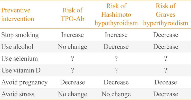
Which preventive interventions can be taken by subjects who are at risk to develop AITD? Table 5 gives an overview of possible interventions that modulate exposure to environmental factors which are likely involved in the immunopathogenesis of AITD [57].
The advice to stop smoking should be given always to every subject. Cessation of smoking will decrease the risk of Graves' hyperthyroidism and even more so of Graves' ophthalmopathy, but the irony is that you lose the protective effect of smoking on Hashimoto's thyroiditis. Moderate use of alcohol seems to protect against both Graves and Hashimoto disease; although, this recommendation will be welcomed by many subjects, drinking alcohol has negative effects as well. Selenium supplementation has been studied extensively in RCTs, but there is still no convincing evidence that increasing selenium intake may prevent a rise of TPO-Ab and thereby the development of subclinical hypothyroidism. Taking 200 µg of selenium daily is unlikely to do harm if baseline serum selenium concentration is lower than 120 µg/L, but carries a risk of type 2 diabetes at higher concentrations. An association between low vitamin D levels and thyroid autoimmunity is likely, but there have been no intervention studies to evaluate whether vitamin D supplementation decreases the risk of thyroid autoimmunity; until such studies have been done, I would not recommend vitamin D for this indication. To avoid pregnancy will indeed decrease the risk of AITD, but that kind of recommendation is not very realistic. And lastly, to avoid stress is likely to diminish the risk of Graves disease, but how do you manage to banish stressful life events and daily hassles from your life?
Taken together, preventive interventions to diminish the risk of AITD are few, not always feasible, and probably of limited efficacy.

Notes

CONFLICTS OF INTEREST: No potential conflict of interest relevant to this article was reported.

References

1. Weetman AP. The immunopathogenesis of chronic autoimmune thyroiditis one century after hashimoto. Eur Thyroid J. 2013; 1:243–250. PMID: 24783026.
crossref
2. Brix TH, Kyvik KO, Christensen K, Hegedus L. Evidence for a major role of heredity in Graves' disease: a population-based study of two Danish twin cohorts. J Clin Endocrinol Metab. 2001; 86:930–934. PMID: 11158069.
crossref
3. Hansen PS, Brix TH, Iachine I, Kyvik KO, Hegedus L. The relative importance of genetic and environmental effects for the early stages of thyroid autoimmunity: a study of healthy Danish twins. Eur J Endocrinol. 2006; 154:29–38. PMID: 16381988.
crossref
4. Brix TH, Hegedus L. Twin studies as a model for exploring the aetiology of autoimmune thyroid disease. Clin Endocrinol (Oxf). 2012; 76:457–464. PMID: 22168537.
crossref
5. Lee HJ, Li CW, Hammerstad SS, Stefan M, Tomer Y. Immunogenetics of autoimmune thyroid diseases: a comprehensive review. J Autoimmun. 2015; 64:82–90. PMID: 26235382.
crossref
6. Villanueva R, Greenberg DA, Davies TF, Tomer Y. Sibling recurrence risk in autoimmune thyroid disease. Thyroid. 2003; 13:761–764. PMID: 14558919.
crossref
7. Strieder TG, Tijssen JG, Wenzel BE, Endert E, Wiersinga WM. Prediction of progression to overt hypothyroidism or hyperthyroidism in female relatives of patients with autoimmune thyroid disease using the Thyroid Events Amsterdam (THEA) score. Arch Intern Med. 2008; 168:1657–1663. PMID: 18695079.
crossref
8. Vanderpump MP, Tunbridge WM, French JM, Appleton D, Bates D, Clark F, et al. The incidence of thyroid disorders in the community: a twenty-year follow-up of the Whickham Survey. Clin Endocrinol (Oxf). 1995; 43:55–68. PMID: 7641412.
crossref
9. Flynn RW, MacDonald TM, Morris AD, Jung RT, Leese GP. The thyroid epidemiology, audit, and research study: thyroid dysfunction in the general population. J Clin Endocrinol Metab. 2004; 89:3879–3884. PMID: 15292321.
crossref
10. Pedersen IB, Knudsen N, Carle A, Vejbjerg P, Jorgensen T, Perrild H, et al. A cautious iodization programme bringing iodine intake to a low recommended level is associated with an increase in the prevalence of thyroid autoantibodies in the population. Clin Endocrinol (Oxf). 2011; 75:120–126. PMID: 21521277.
crossref
11. Pedersen IB, Laurberg P, Knudsen N, Jorgensen T, Perrild H, Ovesen L, et al. An increased incidence of overt hypothyroidism after iodine fortification of salt in Denmark: a prospective population study. J Clin Endocrinol Metab. 2007; 92:3122–3127. PMID: 17504896.
12. Aghini Lombardi F, Fiore E, Tonacchera M, Antonangeli L, Rago T, Frigeri M, et al. The effect of voluntary iodine prophylaxis in a small rural community: the Pescopagano survey 15 years later. J Clin Endocrinol Metab. 2013; 98:1031–1039. PMID: 23436921.
crossref
13. Wiersinga WM. Smoking and thyroid. Clin Endocrinol (Oxf). 2013; 79:145–151. PMID: 23581474.
crossref
14. Strieder TG, Prummel MF, Tijssen JG, Endert E, Wiersinga WM. Risk factors for and prevalence of thyroid disorders in a cross-sectional study among healthy female relatives of patients with autoimmune thyroid disease. Clin Endocrinol (Oxf). 2003; 59:396–401. PMID: 12919165.
crossref
15. Belin RM, Astor BC, Powe NR, Ladenson PW. Smoke exposure is associated with a lower prevalence of serum thyroid autoantibodies and thyrotropin concentration elevation and a higher prevalence of mild thyrotropin concentration suppression in the third National Health and Nutrition Examination Survey (NHANES III). J Clin Endocrinol Metab. 2004; 89:6077–6086. PMID: 15579761.
crossref
16. Asvold BO, Bjoro T, Nilsen TI, Vatten LJ. Tobacco smoking and thyroid function: a population-based study. Arch Intern Med. 2007; 167:1428–1432. PMID: 17620538.
17. Effraimidis G, Tijssen JG, Wiersinga WM. Discontinuation of smoking increases the risk for developing thyroid peroxidase antibodies and/or thyroglobulin antibodies: a prospective study. J Clin Endocrinol Metab. 2009; 94:1324–1328. PMID: 19141579.
crossref
18. Carlé A, Bulow Pedersen I, Knudsen N, Perrild H, Ovesen L, Banke Rasmussen L, et al. Smoking cessation is followed by a sharp but transient rise in the incidence of overt autoimmune hypothyroidism: a population-based, case-control study. Clin Endocrinol (Oxf). 2012; 77:764–772. PMID: 22651374.
19. Effraimidis G, Tijssen JG, Wiersinga WM. Alcohol consumption as a risk factor for autoimmune thyroid disease: a prospective study. Eur Thyroid J. 2012; 1:99–104. PMID: 24783003.
crossref
20. Carlé A, Pedersen IB, Knudsen N, Perrild H, Ovesen L, Rasmussen LB, et al. Moderate alcohol consumption may protect against overt autoimmune hypothyroidism: a population-based case-control study. Eur J Endocrinol. 2012; 167:483–490. PMID: 22802427.
crossref
21. Carlé A, Bulow Pedersen I, Knudsen N, Perrild H, Ovesen L, Rasmussen LB, et al. Graves' hyperthyroidism and moderate alcohol consumption: evidence for disease prevention. Clin Endocrinol (Oxf). 2013; 79:111–119. PMID: 23170908.
crossref
22. Effraimidis G, Strieder TG, Tijssen JG, Wiersinga WM. Natural history of the transition from euthyroidism to overt autoimmune hypo- or hyperthyroidism: a prospective study. Eur J Endocrinol. 2011; 164:107–113. PMID: 20956436.
crossref
23. Gärtner R, Gasnier BC, Dietrich JW, Krebs B, Angstwurm MW. Selenium supplementation in patients with autoimmune thyroiditis decreases thyroid peroxidase antibodies concentrations. J Clin Endocrinol Metab. 2002; 87:1687–1691. PMID: 11932302.
24. Duntas LH, Mantzou E, Koutras DA. Effects of a six month treatment with selenomethionine in patients with autoimmune thyroiditis. Eur J Endocrinol. 2003; 148:389–393. PMID: 12656658.
crossref
25. Turker O, Kumanlioglu K, Karapolat I, Dogan I. Selenium treatment in autoimmune thyroiditis: 9-month follow-up with variable doses. J Endocrinol. 2006; 190:151–156. PMID: 16837619.
crossref
26. Karanikas G, Schuetz M, Kontur S, Duan H, Kommata S, Schoen R, et al. No immunological benefit of selenium in consecutive patients with autoimmune thyroiditis. Thyroid. 2008; 18:7–12. PMID: 18302514.
crossref
27. Nacamulli D, Mian C, Petricca D, Lazzarotto F, Barollo S, Pozza D, et al. Influence of physiological dietary selenium supplementation on the natural course of autoimmune thyroiditis. Clin Endocrinol (Oxf). 2010; 73:535–539. PMID: 20039895.
crossref
28. Eskes SA, Endert E, Fliers E, Birnie E, Hollenbach B, Schomburg L, et al. Selenite supplementation in euthyroid subjects with thyroid peroxidase antibodies. Clin Endocrinol (Oxf). 2014; 80:444–451. PMID: 23844613.
crossref
29. Pilli T, Cantara S, Schomburg L, Cenci V, Cardinale S, Heid EC, et al. IFNγ-inducible chemokines decrease upon selenomethionine supplementation in women with euthyroid autoimmune thyroiditis: comparison between two doses of selenomethionine (80 or 160µg) versus placebo. Eur Thyroid J. 2015; 4:226–233. PMID: 26835425.
30. de Farias CR, Cardoso BR, de Oliveira GM, de Mello Guazzelli IC, Catarino RM, Chammas MC, et al. A randomized-controlled, double-blind study of the impact of selenium supplementation on thyroid autoimmunity and inflammation with focus on the GPx1 genotypes. J Endocrinol Invest. 2015; 38:1065–1074. PMID: 25894865.
crossref
31. Marcocci C, Kahaly GJ, Krassas GE, Bartalena L, Prummel M, Stahl M, et al. Selenium and the course of mild Graves' orbitopathy. N Engl J Med. 2011; 364:1920–1931. PMID: 21591944.
crossref
32. Negro R, Greco G, Mangieri T, Pezzarossa A, Dazzi D, Hassan H. The influence of selenium supplementation on postpartum thyroid status in pregnant women with thyroid peroxidase autoantibodies. J Clin Endocrinol Metab. 2007; 92:1263–1268. PMID: 17284630.
crossref
33. Mao J, Pop VJ, Bath SC, Vader HL, Redman CW, Rayman MP. Effect of low-dose selenium on thyroid autoimmunity and thyroid function in UK pregnant women with mild-to-moderate iodine deficiency. Eur J Nutr. 2016; 55:55–61. PMID: 25524327.
crossref
34. Rayman MP. Selenium and human health. Lancet. 2012; 379:1256–1268. PMID: 22381456.
crossref
35. Wu Q, Rayman MP, Lv H, Schomburg L, Cui B, Gao C, et al. Low population selenium status is associated with increased prevalence of thyroid disease. J Clin Endocrinol Metab. 2015; 100:4037–4047. PMID: 26305620.
crossref
36. Van Belle TL, Gysemans C, Mathieu C. Vitamin D in autoimmune, infectious and allergic diseases: a vital player? Best Pract Res Clin Endocrinol Metab. 2011; 25:617–632. PMID: 21872803.
crossref
37. Wang J, Lv S, Chen G, Gao C, He J, Zhong H, et al. Meta-analysis of the association between vitamin D and autoimmune thyroid disease. Nutrients. 2015; 7:2485–2498. PMID: 25854833.
crossref
38. Choi YM, Kim WG, Kim TY, Bae SJ, Kim HK, Jang EK, et al. Low levels of serum vitamin D3 are associated with autoimmune thyroid disease in pre-menopausal women. Thyroid. 2014; 24:655–661. PMID: 24320141.
crossref
39. Effraimidis G, Badenhoop K, Tijssen JG, Wiersinga WM. Vitamin D deficiency is not associated with early stages of thyroid autoimmunity. Eur J Endocrinol. 2012; 167:43–48. PMID: 22518050.
crossref
40. Feng M, Li H, Chen SF, Li WF, Zhang FB. Polymorphisms in the vitamin D receptor gene and risk of autoimmune thyroid diseases: a meta-analysis. Endocrine. 2013; 43:318–326. PMID: 23065592.
crossref
41. Tomer Y, Davies TF. Infection, thyroid disease, and autoimmunity. Endocr Rev. 1993; 14:107–120. PMID: 8491150.
crossref
42. Wang Z, Zhang Q, Lu J, Jiang F, Zhang H, Gao L, et al. Identification of outer membrane porin f protein of Yersinia enterocolitica recognized by antithyrotopin receptor antibodies in Graves' disease and determination of its epitope using mass spectrometry and bioinformatics tools. J Clin Endocrinol Metab. 2010; 95:4012–4020. PMID: 20484489.
43. Effraimidis G, Tijssen JG, Strieder TG, Wiersinga WM. No causal relationship between Yersinia enterocolitica infection and autoimmune thyroid disease: evidence from a prospective study. Clin Exp Immunol. 2011; 165:38–43. PMID: 21488870.
crossref
44. Brix TH, Hansen PS, Hegedus L, Wenzel BE. Too early to dismiss Yersinia enterocolitica infection in the aetiology of Graves' disease: evidence from a twin case-control study. Clin Endocrinol (Oxf). 2008; 69:491–496. PMID: 18284638.
45. Strieder TG, Wenzel BE, Prummel MF, Tijssen JG, Wiersinga WM. Increased prevalence of antibodies to enteropathogenic Yersinia enterocolitica virulence proteins in relatives of patients with autoimmune thyroid disease. Clin Exp Immunol. 2003; 132:278–282. PMID: 12699417.
crossref
46. Winsa B, Adami HO, Bergstrom R, Gamstedt A, Dahlberg PA, Adamson U, et al. Stressful life events and Graves' disease. Lancet. 1991; 338:1475–1479. PMID: 1683917.
crossref
47. Sonino N, Girelli ME, Boscaro M, Fallo F, Busnardo B, Fava GA. Life events in the pathogenesis of Graves' disease: a controlled study. Acta Endocrinol (Copenh). 1993; 128:293–296. PMID: 8498147.
crossref
48. Kung AW. Life events, daily stresses and coping in patients with Graves' disease. Clin Endocrinol (Oxf). 1995; 42:303–308. PMID: 7758236.
crossref
49. Radosavljevic VR, Jankovic SM, Marinkovic JM. Stressful life events in the pathogenesis of Graves' disease. Eur J Endocrinol. 1996; 134:699–701. PMID: 8766938.
50. Effraimidis G, Tijssen JG, Brosschot JF, Wiersinga WM. Involvement of stress in the pathogenesis of autoimmune thyroid disease: a prospective study. Psychoneuroendocrinology. 2012; 37:1191–1198. PMID: 22226433.
crossref
51. Bülow Pedersen I, Laurberg P, Knudsen N, Jorgensen T, Perrild H, Ovesen L, et al. Lack of association between thyroid autoantibodies and parity in a population study argues against microchimerism as a trigger of thyroid autoimmunity. Eur J Endocrinol. 2006; 154:39–45. PMID: 16381989.
crossref
52. Massoudi MS, Meilahn EN, Orchard TJ, Foley TP Jr, Kuller LH, Costantino JP, et al. Prevalence of thyroid antibodies among healthy middle-aged women. Findings from the thyroid study in healthy women. Ann Epidemiol. 1995; 5:229–233. PMID: 7606312.
53. Frank P, Kay CR. Incidence of thyroid disease associated with oral contraceptives. Br Med J. 1978; 2(2):1531. PMID: 728709.
crossref
54. Vestergaard P, Rejnmark L, Weeke J, Hoeck HC, Nielsen HK, Rungby J, et al. Smoking as a risk factor for Graves' disease, toxic nodular goiter, and autoimmune hypothyroidism. Thyroid. 2002; 12:69–75. PMID: 11838733.
crossref
55. Rotondi M, Pirali B, Lodigiani S, Bray S, Leporati P, Chytiris S, et al. The post partum period and the onset of Graves' disease: an overestimated risk factor. Eur J Endocrinol. 2008; 159:161–165. PMID: 18483014.
crossref
56. Stagnaro-Green A, Schwartz A, Gismondi R, Tinelli A, Mangieri T, Negro R. High rate of persistent hypothyroidism in a large-scale prospective study of postpartum thyroiditis in southern Italy. J Clin Endocrinol Metab. 2011; 96:652–657. PMID: 21190974.
crossref
57. Effraimidis G, Wiersinga WM. Mechanisms in endocrinology: autoimmune thyroid disease: old and new players. Eur J Endocrinol. 2014; 170:R241–R252. PMID: 24609834.
crossref
Table 1

Genetic, Environmental, and Existential Factors in the Immunopathogeneis of Autoimmune Thyroid Disease

enm-31-213-i001
Genetic factors
 Major histocompatibility genes (HLA)
 Immunoregulatory genes (CTLA4, PTPN22, FOXP3, CD25, CD40, FRCL3)
 Thyroid specific genes (TSHR, Tg)
Environmental factors
 Iodine intake
 Smoking
 Alcohol
 Selenium
 Vitamin D
 Infections
 Stress
 Drugs (estrogens)
Existential factors
 Female sex
 Parity
 Age
Table 2

The Predictive THEA Score to Estimate the Risk on Developing Overt Autoimmune Hyperthyroidism or Hypothyroidism in the Next 5 Years

enm-31-213-i002
Variable Score
TSH, mU/L
 <0.4 2
 0.4-2.0 0
 2.1-4.0 2
 4.1-5.7 4
 >5.7 6
TPO-Ab, kU/L
 <100 0
 100-1,000 4
 1,001-10,000 6
 >10,000 12
Family members
 2 Members Graves 1
 2 Members Hashimoto 3
Maximal score 21
THEA score, observed/expected event, %
 0-7 Low 2.7/2.6
 8-10 Medium 14.6/13.1
 11-15 High 27.1/32.9
 16-21 Very high 76.9/59.4

Adapted from Strieder et al. [7].

THEA, Thyroid Events Amsterdam; TSH, thyroid-stimulating hormone; TPO-Ab, thyroid peroxidase antibody.

Table 3

Effect of Selenium Supplementation on TPO antibodies in Eight Randomized Clinical Trials

enm-31-213-i003
Study Selenium preparation Outcome TPO-Ab On L-T4 No. of patients, placebo/selenium Baseline Se, µg/L Baseline TPO-Ab, kU/L
Gartner et al. (2002) [23] Selenite Fall Yes 34/36 69 904
Duntas et al. (2003) [24] SeMet Fall Yes 31/34 75 1,875
Turker et al. (2006) [25] SeMet Fall Yes 40/48 - 803
Karanikas et al. (2008) [26] Selenite No change Yes 18/18 75 524
Nacamulli et al. (2010) [27] Selenite No changea No 30/46 - 172
Eskes et al. (2014) [28] Selenite No change No 30/31 74 1,508
Pilli et al. (2015) [29] SeMet No change No 20/40 82 409
de Farias et al. (2015) [30] SeMet Fall Yes 27/28 37 1,009

TPO-Ab, thyroid peroxidase antibody; Se, selenium; Selenite, sodium selenite; SeMet, selenomethionine.

aFall at 1 year.

Table 4

Low Selenium Intake Is Associated with High Prevalence of Hashimoto Thyroiditis: A Population-Based Study in China

enm-31-213-i004
Variable County with adequate Se intake
(n=3,038)
County with low Se intake
(n=3,114)
P value
Serum Se, µg/L, median (IQR) 104 (80-136) 57 (39-82) <0.05
Hypothyroidism, % 2.0 4.2 <0.001
Subclinical hypothyroidism, % 11.7 21.4 <0.001
Autoimmune thyroid, % 2.2 3.4 0.007
Subclinical hyperthyroidism, % 1.3 0.6 0.003
Hyperthyroidism, % 1.3 1.7 NS
Graves disease, % 0.5 0.7 NS
Enlarged thyroid, % 7.7 12.3 0.001
Nodules, % 7.6 7.2 NS

Adapted from Wu et al. [35].

Se, selenium; IQR, interquartile range; NS, not significant.

Table 5

Modulation of Exposure to Environmental Factors in Order to Decrease the Risk of Developing Autoimmune Thyroid Disease

enm-31-213-i005
Preventive intervention Risk of TPO-Ab Risk of Hashimoto hypothyroidism Risk of Graves hyperthyroidism
Stop smoking Increase Increase Decrease
Use alcohol No change Decrease Decrease
Use selenium ? ? ?
Use vitamin D ? ? ?
Avoid pregnancy Decrease Decrease Decease
Avoid stress No change No change Decrease

Adapted from Effraimidis et al. [57].

TPO-Ab, thyroid peroxidase antibody.

TOOLS
Similar articles