

 PDF
PDF ePub
ePub Citation
 Print
Print


INTRODUCTION
Breast cancer is the most common female malignancy and the second leading cause of cancer-related death in the United States. Among patients who die from breast cancer, more than 70% suffer from bone metastasis, which is often accompanied by severe bone pain, fracture, and potentially lethal complications such as hypercalcemia [1234]. Currently, although anti-osteolytic agents (such as bisphosphonate and receptor activator of nuclear factor kappa-β ligand [RANKL] antibody denosumab) [567], radiotherapy, and chemotherapy can reduce morbidity associated with bone metastasis, these treatments often do not significantly extend the survival time of the patients or provide a cure [89], as metastatic cancers often acquire resistance to these treatments. More effective therapies are needed to improve the clinical outcome for stage IV breast cancer patients with bone metastasis. Tumor-stromal interaction plays a major role in promoting bone metastasis of breast cancer [4]. The bone microenvironment mainly includes osteoblast lineage cells, osteoclasts, hematopoietic cells, and additional stromal cell types residing in the bone marrow (BM). Formation of bone metastasis is the result of complicated interactions between tumor cells and various stromal cells in bone, leading to the initial survival of cancer cells in the bone microenvironment, activation from dormancy or indolent growth, and expansion of overt osteolytic lesions. Understanding the molecular mechanism of tumor-stromal interactions in bone metastasis is crucial for the improved early detection of bone metastasis, as well as more effective therapies to prevent or slow down the development of bone metastasis.
STROMAL CELLS IN THE BONE MICROENVIRONMENT
As a crucial organ to foster hematopoiesis and osteogenesis in healthy individuals, bone represents a biologically highly active microenvironment containing various stromal niches to regulate the dynamic balance of stem, progenitor and mature cells of different lineages [1011]. Recent studies from the field of hematopoiesis indicate the existence of two major niches in the BM: the osteoblastic niche and the perivascular niche. Within these niches, two major cell lineages derived from hematopoietic stem cells (HSCs) and mesenchymal stem/stromal cells (MSCs) have complex interactions with each other to sustain normal hematopoiesis and osteogenesis [111213]. Located at the inner surface of the bone cavity, the osteoblastic niche has been previously reported to mainly accommodate long-term quiescent HSCs, although recent findings revealed it as the primary site for early B-cell progenitors and certain lymphoid progenitors [12141516]. In contrast, recent studies suggested that HSCs are mostly localized in the perivascular niche where the endothelial cells, C-X-C motif chemokine 12 (CXCL12)-abundant reticular (CAR) cells and MSCs regulate the HSCs through a series of growth factors, cytokines and chemokines such as stem cell factor (SCF), CXCL12, and angiopoietin-1 [12]. In particular, BM-MSCs are capable of generating osteoprogenitor cells to form the osteoblastic niche, and releasing CXCL12, thrombopoietin and other factors to maintain HSC self-renewal and proliferation. In addition to maintaining healthy bone development, the bone niche supplies immune cells and tissue progenitor cells reconstitute the peripheral immune system and contribute to tissue repair and regeneration [171819]. The osteoblastic and the perivascular niches have both been reported to impact metastatic survival and tumor cell proliferation during bone metastasis. Direct competition for the osteoblastic niche has been observed between HSCs and metastatic cancer cells [20]. The perivascular niche has been characterized as an alternate site for bone metastatic colonization [212223].
Osteoblasts are differentiated from MSCs [24]. Together with osteocytes that have been terminally differentiated from osteoblasts, these cells are the major cell types with bone building functions. Through depositing cross-linked collagen, calcium and other mineral substrates, osteoblast cells are responsible for building up the hard yet elastic bone matrix [25]. Osteoblasts also play a significant role in maintaining bone homeostasis, as osteoblasts secrete cytokine RANKL to promote the maturation of the bone degrading osteoclasts [26]. Recent studies have demonstrated that disseminated tumor cells (DTCs) in the bone microenvironment usually engage osteoblasts to survive and form proliferating colonies. DTCs have been shown to compete with HSCs for occupancy in the osteoblast niches [20]. In breast cancer bone metastasis, tumor cells use heterotypic cadherin interactions with osteogenic cells to activate prosurvival mammalian target of rapamycin-Akt signaling [27].
The bone resorbing osteoclast is another major stromal cell type in bone that play an important role in physiological bone remodeling [2829], and in pathological conditions such as Paget's disease and lytic bone metastasis [4]. Osteoclast differentiation is crucially dependent on macrophage colony-stimulating factor (M-CSF) and RANKL [2829] and are additionally controlled by other growth factors and cytokines [2829]. As the only cell type in human body that is capable of bone degradation, osteoclasts has been the focus in the study of tumor-stromal interactions in bone metastasis, and the development of osteoclast-targeting treatments for bone metastasis [30].
Other bone stromal cells that have been implicated in the development of bone metastasis include CD4+ T cells, myeloid-derived suppressor cells (MDSCs), Tregs, and Dendritic cells. Contrary to the common perception of the anti-tumor effects of T cells, CD4+ T cells have been demonstrated to be part of the pre-metastatic niche in bone, and activate bone remodeling by secreting RANKL [31]. Inflammatory molecule prostaglandin E2 released from breast tumor cells have also been reported to recruit Tregs to establish a pro-metastatic niche in bone [32]. Furthermore, plasmocytoid dendritic cells have been shown to recruit MDSCs and Tregs and inhibit the cytotoxicity of CD8+ T cells to promote bone metastasis [33].
TUMOR-STROMAL INTERACTIONS IN BONE METASTASIS
Bone tissue constantly undergoes dynamic remodeling mediated by the balanced activity of osteoclasts and osteoblasts. Metastatic cancer cells often exploit the normal bone homeostatic process and tip the equilibrium toward either hyperactive bone lysis or bone growth to facilitate the formation of bone metastasis. The proclivity of breast cancer in forming osteolytic bone metastasis has been frequently cited as the classic example of "seed and soil" interactions between tumor and stroma in metastasis. A "vicious cycle" of molecular crosstalk between tumor cells and the bone microenvironment often takes place in osteolytic bone metastasis whereby tumor cell-produced factors stimulate osteoclast maturation or activity, leading to extensive degradation of the bone matrix and the release of bone-derived factors that further enhance tumor growth. A major effort of our research is devoted to identifying molecular mediators used by tumor cells to engage various stromal cell types in the bone microenvironment to promote the initiation and progression of bone metastasis.
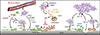
Most bone relapses of breast cancer occur many years after the initial treatment of primary tumors. The molecular basis for the activation of dormant bone micrometastasis to life-threatening overt metastasis remains largely unknown, in large part due to the lack of appropriate animal models that closely recapitulate the process. We developed a novel mouse model to mimic the activation of indolent micrometastases to aggressive lesions. In this model, the single cell progeny clone number 6 (SCP6) single-cell derived clone of the MDA-MB-231 breast cancer cell line was known to have no basal bone metastatic ability after intracardiac injection into nude mice recipients [34]. Interestingly, a small number of mice eventually developed osteolytic bone lesions after more than 6 months of apparent bone metastasis free survival, suggesting that certain genetic/epigenetic changes occurs in the DTCs that endowed them with bone metastatic ability. Careful gene expression profiling analysis and functional studies led to the identification and validation of vascular cell adhesion molecule 1 (VCAM1) as a crucial functional driver of conversion from indolent micrometastases to overt osteolytic bone metastasis [35]. Mechanistically, we determined that tumor-derived soluble VCAM1 serves as a chemoattractant to recruit circulating monocytic precursors of osteoclasts. By interacting with its cognate receptor, α4β1 integrin, VCAM1 also promotes the adhesion of preosteoclasts to the surface of tumor cells, leading to cell fusion and differentiation of pre-osteoclasts and initiation of bone destruction (Fig. 1).
To systematically identify tumor-derived factors that promote bone metastasis, we developed an in vivo selection strategy to isolate bone-metastatic breast cancer variants [34]. The MDA-MB-231 cell line contains a heterogeneous population of cancer cells based on morphological and gene expression analysis. When the parental cell line was injected into nude mice via the left cardiac ventricle to form bone metastasis, about 20% to 30% of mice developed osteolytic bone lesions. More than half of the sublines of cancer cells isolated from these lesions displayed dramatically increased ability to metastasize to bone, while some sublines displayed mildly or no increase of bone metastatic ability. These isogenic sublines with differential bone metastatic ability provided an ideal cohort to identify candidate bone metastasis genes based on gene expression profiling. Genes in the bone metastasis expression signature included previously reported bone metastasis genes, such as C-X-C chemokine receptor type 4 (CXCR4) [36], but also contains many novel candidate metastasis genes that were subsequently validated in follow-up studies, including interleukin 11 (IL-11), osteopontin, connective tissue growth factor (CTGF), Jagged1, matrix metalloproteinase-1 (MMP1), ADAM metallopeptidase with thrombospondin type 1 motif, 1 (ADAMTS1), and chemokine (C-C motif) ligand 2 (CCL2) [34373839]. Functional characterization of candidate bone metastasis genes revealed novel mechanisms of tumor-stromal interactions. For example, we showed that two metalloproteases, MMP1 and ADAMTS1, perform important signaling functions in osteoclast differentiation through activating a paracrine cascade mediated by three different cell types [38]. MMP1 and ADAMTS1 proteolytically cleave the membrane-bound epidermal growth factor (EGF) family ligands, including heparinbinding epidermal growth factor-like growth factor (HB-EGF) and amphiregulin, which activate epidermal growth factor receptor (EGFR) signaling in osteoblasts, leading to reduced expression of osteoprotegerin, the decoy receptor and antagonist of RANKL. Increased RANKL activity promotes osteoclast differentiation and osteolytic bone metastasis (Fig. 1).
It is believed that growth factors embedded in bone matrix are released during bone destruction and further stimulate the malignancy of cancer cells, forming a "vicious cycle" in bone metastasis. Among the bone-derived growth factors, we are particularly interested in the role of transforming growth factor β (TGF-β) since it is one of the most abundant bone-embedded growth factors. Furthermore, many of the bone metastasis genes are direct transcriptional targets of TGF-β. We first used genetic, pharmacological and advanced imaging approaches to demonstrate that TGF-β is released from the bone during bone destruction and further promotes tumor malignancy [40]. Using a MDA-MB-231 cell line engineered to have conditional Smad4 expression and also contain a dual luciferase report system for imaging TGF-β signaling activity (using firefly luciferase driven by Smad binding elements) and tumor burden (using cytomegalovirus promoter driven Renilla luciferase), we explored the temporal-spatial dynamics and requirement of TGF-β signaling in bone metastasis. We showed that TGF-β signaling activity was dramatically elevated in osteolytic bone lesions, and such activation was inhibited when the mice are treated with bisphosphonates to reduce bone lysis. This result indicated that bone is indeed a major source of TGF-β during bone metastasis. Importantly, both genetic (using Tet-off expression control of Smad4) and pharmacological (using TGF-β receptor kinase inhibitor treatment) inhibition of TGF-β signaling in mice dramatically reduce the development of bone metastasis [40].
We subsequently identified Jagged1 as an important TGF-β downstream target with a crucial role in engaging bone stromal cells in osteolytic metastasis [39]. Tumor-derived Jagged1 activates Notch signaling in osteoblasts to increase the expression of IL-6, which feeds back to tumor cells to stimulate their growth. In parallel, Jagged1 also directly activates osteoclast differentiation for bone destruction (Fig. 1). Thus, two developmentally conserved pathways, TGF-β and Notch, converge to constitute a vicious cycle that may account for the frequent bone metastasis of breast cancer. This research has led to our current collaborative effort with Amgen (Thousand Oaks, CA, USA) to develop humanized, Jagged1-blocking antibodies, which are showing excellent efficacy in pre-clinical testing.
Turning our attention to bone stromal cells, we delineated a micro RNA (miRNA) regulatory network [41] that controls the activation of osteoclasts by RANKL and tumor-derived factors, such as soluble intercellular adhesion molecule 1 (ICAM1) [41] and CCL2 [37]. Osteoclast miRNAs down-regulated during osteoclastogenesis are potent inhibitors of bone resorption and osteolytic bone metastasis, while up-regulated miRNAs, such as miR-16 and miR-378, can be detected in circulation as biomarkers of bone metastasis (Fig. 1) [41]. Remarkably, treatment of mice with miRNAs that are down-regulated during bone metastasis led to significant inhibition of bone metastasis development, suggesting that miRNAs can be developed as novel therapeutic agents that target bone stromal cells to reduce bone metastasis development.
CONCLUSIONS AND FUTURE PERSPECTIVES
The study of bone metastasis has produced substantial new insights into the intricate cross-talk between metastatic cancer cells and bone stromal cells, particularly osteoblasts and osteoclasts. These findings have resulted in the successful development of bisphosphonates and denosumab (RANKL-neutralizing antibody) as some of the first U.S. Food and Drug Administration-approved stroma-targeting treatments for metastatic cancer. However, such treatments are generally not curative, and simply reduce skeletal-related events (bone fracture, bone pain, etc.) without improving the overall survival of patients. Interestingly, adjuvant treatment of bisphosphonates improved the survival of post-menopausal breast cancer patients [42], suggesting early application of anti-metastasis treatments may have better outcome than in late-stage diseases. With rapid advance in the field of bone metastasis research, it is likely that several new bone metastasis targeting agents such as Jagged1 neutralizing antibody may become available soon. It will be imperative to test the potential synergistic effect of combing different agents with distinct targeting mechanisms. It is possible that stroma-targeting treatment may enhance the efficacy of traditional chemotherapy and radiation therapy, which are commonly used to control bone metastasis, as well as the newly developed immune checkpoint therapy. As bone metastases are usually more refractory to various cancer treatments than the primary tumor, understanding the molecular mechanism of treatment resistance in bone metastasis may further provide a promising avenue of further investigation that may significantly improve the outcome of patients with bone metastasis.
 XML Download