Abstract
Background
Stress affects body weight and food intake, but the underlying mechanisms are not well understood.
Methods
We evaluated the changes in body weight and food intake of ICR male mice subjected to daily 2 hours restraint stress for 15 days. Hypothalamic gene expression profiling was analyzed by cDNA microarray.
Results
Daily body weight and food intake measurements revealed that both parameters decreased rapidly after initiating daily restraint stress. Body weights of stressed mice then remained significantly lower than the control body weights, even though food intake slowly recovered to 90% of the control intake at the end of the experiment. cDNA microarray analysis revealed that chronic restraint stress affects the expression of hypothalamic genes possibly related to body weight control. Since decreases of daily food intake and body weight were remarkable in days 1 to 4 of restraint, we examined the expression of food intake-related genes in the hypothalamus. During these periods, the expressions of ghrelin and pro-opiomelanocortin mRNA were significantly changed in mice undergoing restraint stress. Moreover, daily serum corticosterone levels gradually increased, while leptin levels significantly decreased.
Conclusion
The present study demonstrates that restraint stress affects body weight and food intake by initially modifying canonical food intake-related genes and then later modifying other genes involved in energy metabolism. These genetic changes appear to be mediated, at least in part, by corticosterone.
Stress is well known to change body weight and food intake in animal models. Of the various stress models available for the study of the effects of stress, the restraint stress model is most commonly employed, as it effectively mimics potent physical and psychological stress [1]. The restraint stress model has also been used as an animal model of depression and anorexia nervosa. Thus, many studies have shown that restraint stress suppresses body weight gain and food intake in rodents [2,3].
The central regulation of body weight and food intake occurs in the hypothalamus, which contains multiple neuronal systems that play important roles in the regulation of energy homeostasis [4]. These systems involve the interaction of multiple neuropeptides. Food intake reflects a functional balance between hypothalamic orexigenic peptides (such as neuropeptide Y [NPY] and agouti-related protein [AgRP]) and anorexigenic peptides (such as pro-opiomelanocortin [POMC] and cocaine- and amphetamine-regulated transcript [CART]) [5]. In addition, ghrelin, a peptide that is predominantly produced by the stomach, is also expressed by the hypothalamus and regulates growth hormone secretion, food intake, and energy homeostasis [6,7]. Another factor that regulates food intake and energy homeostasis is leptin, an anorexigenic hormone secreted by adipose tissue [8]. Leptin is well known for its critical role in the regulation of food intake in adult mammals. Furthermore, leptin participates in the control of several neuroendocrine functions, including those of the hypothalamicpituitary-adrenal (HPA) axis. In response to the nutritional status and energy storage levels, leptin signals hypothalamic feeding centers by controlling the expression and release of orexigenic and anorexigenic neuropeptides [9,10].
Chronic stress increases serum corticosterone levels. However, the effects of chronic stress-induced elevated corticosterone on food intake and body weight are not clear [11]. Furthermore, the precise mechanism by which stress affects energy metabolism as well as food intake and body weight control is not well understood, especially at the hypothalamic gene expression level. In this study, to identify the central genes that regulate body weight and food intake and to characterize the molecular mechanisms involved, we extensively analyzed the hypothalamic gene expression profiles of chronically restraint stressed mice using large-scale cDNA microarray analysis.
Male 7-week-old ICR mice were purchased from Central Laboratory Animal Inc. (Seoul, Korea) and housed individually in clear plastic cages in a temperature- and humidity-controlled environment under a 12 hours light/dark cycle (light on at 0600 hour) with free access to lab chow and water. The experiments were performed after the animals had been habituated to the experimental environment for 1 week. The mice were divided into two weight-matched (31 to 33 g) groups, controls, and stressed mice. The stressed mice were exposed daily for 15 days to 2 hours of restraint (0930 to 1130 hours) in an acrylic cylindrical animal restrainer (Φ25×[H] 85 mm, Daejong Instrument Industry, Seoul, Korea) with holes that permit the restrainer to be adjusted according to the size of the subject. The restrainer allows unlimited breathing but restricts the movement of the limbs. After being restrained, the mice were returned to their home cage and given food and water ad libitum. The food consumption and body weight of the mice were monitored daily (0830 to 0900 hours). All animal procedures adhered to the Animal Care and Use Guidelines of Gyeongsang National University (Approval No., GLA-060502-M0002 and GLA-070802-M0035).
One day after stress ended, days 1 to 5 or day 16 (depending on the experiment), the animals were sacrificed and their hypothalami were rapidly extracted (0930 to 1130 hours). Total RNA was isolated using TRIzol reagent (Invitrogen, Carlsbad, CA, USA). The purity and quantity of the RNAs were assess ed by spectrophotometry.
Gene expression analysis was conducted on the day 16 hypothalamic mRNAs using the Agilent Mouse oligo microarray kit (Digital Genomics, Seoul, Korea). The scanned images were analyzed with GenePix Pro 6.0 software (Axon Instruments, Union City, CA, USA) to obtain gene expression ratios. The transformed data were normalized by LOWESS regression and analyzed using GeneSpring GX 7.3 software program (Agilent Technologies Inc., Santa Clara, CA, USA).
The elevated plus maze (EPM) has two open arms and two closed arms (30×7 cm each) and a connecting central platform (7×7 cm) mounted 50 cm above the floor. Tested mice were placed in the center of the maze facing the open arm, and behavior was recorded for 5 minutes. Arm entry was scored if a mouse moved into the arm.
To measure the basal levels of serum corticosterone and leptin, blood was collected the morning after stress via decapitation. Serum corticosterone and leptin concentrations were determined using an EIA kit (Assay Designs Inc., Ann Arbor, MI, USA) and an ELISA kit (Millpore, St Charles, MO, USA), respectively.
For the day 0 to 4 samples of 16 hypothalamic mRNAs, a M-MLV RT kit (Promega, Madison, WI, USA) was used to convert the RNAs (1 µg) to cDNAs. The ghrelin and POMC mRNA levels were then determined using a real-time polymerase chain reaction (PCR) kit (LightCycler FastStart DNA Master SYBR Green I, Roche Applied Science, Mannheim, Germany) with the following primers: ghrelin (292 bp) forward, 5'-CAGTTTGCTGCTACTCAG-3', reverse, 5'-GATATCCTGAAGAAACTTCC-3'; POMC (497 bp) forward, 5'-ATGCCGAGATTCTGCTAC-3', reverse, 5'-AGCTCCCTCTTGAACTCT-3'; glyceraldehyde-3-phosphate dehydrogenase (GAPDH; 172 bp) forward, 5'-TGCCGCCTGGAGAAACCTGC-3', reverse, 5'-TGAGAGCAATGCCAGCCCCA-3'. The hydroxysteroid (17-β) dehydrogenase 1 (Hsd17b1), cytochrome P450, family 11, subfamily a, polypeptide 1 (Cyp11a1), glycoprotein hormones, α subunit (Cga), and growth hormone (Gh) mRNA levels were then determined using a conventional PCR with the following primers: Hsd17b1 (370 bp) forward, 5'-ACTACCTGCGTGGTTATGAG-3', reverse, 5'-TGGTAACATGAATTGTCCTG-3'; Cyp11a1 (375 bp) forward, 5'-CCAAGATGGTACAGTTGGTT-3', reverse, 5'-CATCACGGAGATTTTGAACT-3'; Cga (317 bp) forward, 5'-AGCTAGGAGCCCCCATCTAC-3', reverse, 5'-GCGTCAGAAGTCTGGTAGGG-3'; Gh (255 bp) forward, 5'-TTCTGCTTCTCAGAGACCAT-3', reverse, 5'-TCATAGGTTTGCTTGAGGAT-3'. The expression levels of each mRNA are presented throughout as arbitrary units.
Real-time PCR data were analyzed by LightCycler software version 4.0 (LightCycler 2.0 Instrument, Roche Applied Science). Conventional PCR data were analyzed by Gel Doc (Bio-Rad, Hercules, CA, USA) and Quantity One version 4.6.3. Messenger RNA levels were normalized to the levels of the GAPDH reference gene. EPM test were analyzed using a computerized video-tracking system (EthoVision version 3.0, Noldus Information Technology, Wageningen, the Netherlands). Statistical analysis were performed using Student unpaired t test and one or two-way analysis of variance (GraphPad Prism, La Jolla, CA, USA). All data are shown as mean±SE.
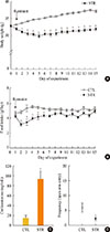
The effects of daily restraint stress for 15 days on body weight and food intake shown in Fig. 1. While the body weights of the control mice gradually increased over the 15 day experiment, the body weights of the stressed mice dropped sharply during the first 5 days. As a result, the stressed mice had significantly lower body weights than the control mice during the entire experimental period (Fig. 1A).
The total food intake of the stressed mice also decreased markedly during the first few days of the experiment. The daily food intake of the stressed mice then gradually recovered substantially. However, while food intake of the stressed mice was almost 90% of control intake by day 7, it remained significantly lower at nearly every time point (Fig. 1B).
Serum corticosterone levels were measured on day 16 (without stress) and were significantly higher in the stressed mice (Fig. 1C). In addition, we measured anxiety levels using the EPM test. The frequency of entries in open arm was significantly lower in the stressed group (Fig. 1D). However, the frequency of entries into the closed arm tended to be increased compared with control group, although the difference was not significant (data not shown).
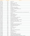
The restraint stressed or control mice (n=10 in each group) were sacrificed on day 16 without stress, and their hypothalamic mRNAs were subjected to large-scale cDNA microarray analysis. Among the 20,868 detected genes, 42 genes showed a significant greater than 2.0-fold increase or 0.5-fold decrease in expression in the stressed mice (Table 1). To confirm the microarray data using conventional PCR analysis, we randomly selected four genes, two that were up-regulated and two that were down-regulated. The Hsd17b1 and Cyp11a1 mRNA levels were significantly increased, while the Cga and Gh mRNA levels showed a significant decrease (Fig. 2).
Chronic restraint stress reduced body weight and food intake. Particularly, in days 1 to 4 of restraint, food intake and body weight were dramatically decreased. Thus, we analyzed the expression of canonical food intake-related genes in this period. To determine the expression of hypothalamic neuropeptides known to be involved in energy homeostasis, such as ghrelin and POMC, the hypothalamic mRNAs from the group of mice sacrificed on each of the 4 days after initiating restraint stress were subjected to real-time PCR analysis. On day 2 of restraint stress, the ghrelin mRNA levels showed a significant decrease, while the POMC mRNA levels were significantly elevated (Fig. 3A, B).
To elucidate the changes of food intake-related hormones levels after stress, we collected blood from the animals daily and measured serum corticosterone and leptin levels. Restraint stress showed gradually increasing serum corticosterone levels (Fig. 3C) and significantly decreased leptin levels on days 2 to 4 of restraint (Fig. 3D).
In the present study, we investigated the effects of restraint stress on body weight, food intake, and hypothalamic gene expression levels in mice. Several studies have demonstrated that chronic exposure to restraint stress reduces the body weight and food intake of rodents [12-14]. However, the mechanisms underlying these restraint-induced changes in body weight and food intake remain to be elucidated.
Our results here showed that restraint stress rapidly induces a marked decrease in body weight that may be due to a reduction of food intake. However, while food intake recovered to 90% of control intake, this was not matched by an equivalent recovery in body weight for the duration of the exposure to restraint. The stress-induced decrease in body weight may be due initially to an early decrease in food intake but then may be subsequently maintained by increases in energy expenditure and body temperature during restraint [15]. Especially, a previous report has also shown that rats chronically exposed to restraint showed rapid weight loss that did not recover even after removal of the stress [13]. Moreover, this study also showed that, while exposure to restraint stress significantly lowered food intake, once the stress ended, the food intake of the stressed group returned to the level of the control group; there was no attempt to overeat to compensate for the energy deficiency experienced during the restraint period [13]. This may be because stress somehow modifies the pathways that would normally sense and respond to a reduction in weight. It has been reported that stress-related pathways, once activated, act in opposition to the mechanisms that would normally promote the recovery of weight to normal levels [16].
Increased serum corticosterone levels are consistent with the suggestion that physiological responses to repeated stress are associated with the activation of the HPA axis [17]. Also, we showed increasing anxiety levels in the stressed mice; chronic stress has been shown to increase anxiety and depression-like behavior in animal models [18,19]. Consequently, these results indicate that chronic restraint stress changes physical and psychological responses.
To determine whether chronic restraint stress affects hypothalamic gene expression in the mice, we subjected the hypothalami of mice that had been exposed to 2 hours of restraint stress daily for 15 consecutive days to cDNA microarray analysis. Many of the genes that showed stress-related changes in expression were related to body weight control. Thus, these genes such as Gh, Prolactin, Cga, STEAP family member 4, Hsd17b1, Cyp11a1, adipsin, and trehalase (see the Genbank_Acc. No.) may participate in the chronic restraint stress-induced reduction of body weight, although this notion remains to be tested (Table 1).
Supporting the possible involvement of metabolism-related genes, a recent study showed that restraint stress affects lipid metabolism. In that study, rats exposed to acute or chronic restraint stress show remarkable changes in plasma lipid and lipoprotein levels; plasma fatty acid, glycerol, and cholesterol levels are increased, while plasma triacylglyceride levels are decreased [20]. Supporting this notion is the finding that chronic restraint stress increases serum corticosterone levels, which may stimulate the catabolism of skeletal muscle proteins, which in turn may, at least in part, lead to body weight loss [21]. Recently, psychological stress has been shown to attenuate body size and lean body mass by reducing muscle mass [22].
The mice that were exposed to chronic restraint stress for 15 days showed sustained reductions in body weight and food intake. As the initial dramatic decreases in daily food intake and body weight were observed in days 1 to 4 of restraint, we hypothesized that canonical food intake-related genes may only participate in this period.
Thus, we analyzed the expression of food intake-related genes, such as NPY, AgRP, POMC, CART, ghrelin, corticotropin-releasing factor (CRF), CRF receptors, leptin receptor, insulin receptor, and melanocortin receptor, using real-time PCR. Only the hypothalamic mRNA expression of the ghrelin and POMC showed a significant decrease and increase on day 2, respectively. It has been shown that ghrelin is an orexigenic factor, as the central administration of ghrelin strongly stimulates food intake and increases body weight [23]. Moreover, when ghrelin is injected in an intracerebroventricular manner, NPY, and AgRP mRNA expressions are increased in the arcuate nucleus (Arc) [24]. In relation to the latter observation, hypothalamic ghrelin neurons are located within the Arc and innervate NPY/AgRP neurons [25]. Thus, it seems that the orexigenic effect of ghrelin is dependent on NPY/AgRP neurons. Moreover, NPY/AgRP neurons innervate POMC/α-melanocyte-stimulating hormone (α-MSH) neurons, indicating that the melanocortin system also seems to be involved in the action of ghrelin [26]. It has been suggested that POMC neurons act anorexigenically by producing and releasing α-MSH, a peptide that activates melanocortin-3, -4 receptors and inhibits food intake [27]. That we observed decreased ghrelin and increased POMC mRNAs early after restraint stress initiation suggests that these proteins may be responsible, at least in part, for the initial weight loss observed after restraint stress induction.
We also observed that the serum corticosterone and leptin levels increased and decreased, respectively, in the 4 days after restraint stress was initiated. Serum leptin levels were decreased from day 2. In another report, restraint stress decreased serum leptin levels, which were sustained even after restraint stress was eliminated [28]. Sustained reduced leptin levels may recover food intake, as shown in our results. During a period of chronic restraint stress, despite the nearly recovered food intake, discrepancy in body weight between stressed and control mice was not reduced. This continued discrepancy of body weight may be possibly due to the action of increasing corticosterone levels. Glucocorticoids have a broad range of activity that affects the expression and regulation of genes throughout the body; these glucocorticoid-mediated effects lead to changes in the energy and metabolism requirements of the organism [29]. In our study, initial loss of body weight might be caused by reduction of food intake after stress, and this finding is well match with other reports [12,30]. According to several studies, exposure to chronic stress in rats resulted in an increase in basal corticosterone levels [31,32]. These results probably reflect a modified sensitivity to the negative feedback effects of circulating glucocorticoid [33]. In addition, food intake and many metabolic processes are mediated by glucocorticoids. Thus, chronic stress has been related to changes in body weight and physiology of different organs [15,32]. In our study, serum corticosterone levels were increased by repeated restraint stress. Thus, we suggest that the increased serum corticosterone levels after restraint stress could be due to a continuous stress state. This increased serum corticosterone level might affect discrepancies in body weight and food intake recovery in a direct or indirect manner. Daily increased pattern of serum corticosterone levels may affect the serum leptin levels. Although decreased serum leptin levels in early days of stress seemed to have a role in the recovered food intake, the precise role of serum leptin and the correlation of leptin with corticosterone needs further evaluation.
In summary, restraint stress affects the body weight and food intake in mice. Chronic restraint stress-induced reduction of body weight is caused by reduction of initial daily food intake through modification of canonical food intake-related genes. However, chronic restraint stress-induced sustained discrepancy of body weight without reduction of food intake may be due to expression of other genes related to body weight control and regulation of stress response through corticosterone.
Figures and Tables
Fig. 1
Effects of restraint stress on body weight, food intake, serum corticosterone, and anxiety level. (A) Daily body weight and (B) food intake of mice exposed daily to 2 hours of restraint for 15 consecutive days. (C) Serum corticosterone levels were significantly increased in stressed mice (STR) at the end of the restraint stress period. (D) Stressed mice showed a significant reduction in frequency of open arm entry in the elevated plus maze test. Statistical differences were evaluated by (A, B) two-way analysis of variance and (C, D) Student unpaired t test. Data are presented as mean±SE. aP<0.05; bP<0.01 vs. control mice (CTL) (n=10 in each group).

Fig. 2
Reverse transcriptase polymerase chain reaction analysis of the altered gene expression identified from microarray analysis. The levels of hydroxysteroid (17-β) dehydrogenase 1 (Hsd17b1) and cytochrome P450, family 11, subfamily a, polypeptide 1 (Cyp11a1) mRNA were increased in stressed mice (STR), while glycoprotein hormones, α subunit (Cga) and growth hormone (Gh) mRNA levels were decreased. Statistical difference was evaluated by Student unpaired t test. Data are presented as mean±SE. aP<0.05; bP<0.01 vs. control mice (CTL) (n=6 in each group).

Fig. 3
Effects of restraint stress on hypothalamic gene expression and serum hormone levels. (A) Hypothalamic mRNA expression of ghrelin and (B) pro-opiomelanocortin (POMC) in mice exposed daily to 2 hours of restraint for 4 days. (C) Serum corticosterone and (D) leptin levels for mice exposed daily to 2 hours of restraint for 4 days. Statistical differences were evaluated by one-way analysis of variance and Dunnett t test. Data are presented as mean±SE. aP<0.05; bP<0.01 vs. control mice (n=6 in each group).

ACKNOWLEDGMENTS
This work was supported by the Korea Science and Engineering Foundation (KOSEF) for the Basic Research Program (R01-2006-000-10259-0) and partially supported by the Basic Science Research Program through the National Research Foundation (NRF) of Korea, funded by the Ministry of Education, Science and Technology (R13-2005-012-02001-0).
References
1. Glavin GB, Pare WP, Sandbak T, Bakke HK, Murison R. Restraint stress in biomedical research: an update. Neurosci Biobehav Rev. 1994; 18:223–249.
2. Krahn DD, Gosnell BA, Majchrzak MJ. The anorectic effects of CRH and restraint stress decrease with repeated exposures. Biol Psychiatry. 1990; 27:1094–1102.
3. Dallman MF, Akana SF, Scribner KA, Bradbury MJ, Walker CD, Strack AM, Cascio CS. Stress, feedback and facilitation in the hypothalamo-pituitary-adrenal axis. J Neuroendocrinol. 1992; 4:517–526.
4. Woods SC, Seeley RJ, Porte D Jr, Schwartz MW. Signals that regulate food intake and energy homeostasis. Science. 1998; 280:1378–1383.
5. Korner J, Savontaus E, Chua SC Jr, Leibel RL, Wardlaw SL. Leptin regulation of Agrp and Npy mRNA in the rat hypothalamus. J Neuroendocrinol. 2001; 13:959–966.
6. Tschop M, Smiley DL, Heiman ML. Ghrelin induces adiposity in rodents. Nature. 2000; 407:908–913.
7. van der Lely AJ, Tschop M, Heiman ML, Ghigo E. Biological, physiological, pathophysiological, and pharmacological aspects of ghrelin. Endocr Rev. 2004; 25:426–457.
8. Proulx K, Richard D, Walker CD. Leptin regulates appetite-related neuropeptides in the hypothalamus of developing rats without affecting food intake. Endocrinology. 2002; 143:4683–4692.
9. Ahima RS, Prabakaran D, Mantzoros C, Qu D, Lowell B, Maratos-Flier E, Flier JS. Role of leptin in the neuroendocrine response to fasting. Nature. 1996; 382:250–252.
10. Elmquist JK, Maratos-Flier E, Saper CB, Flier JS. Unraveling the central nervous system pathways underlying responses to leptin. Nat Neurosci. 1998; 1:445–450.
11. Smagin GN, Howell LA, Redmann S Jr, Ryan DH, Harris RB. Prevention of stress-induced weight loss by third ventricle CRF receptor antagonist. Am J Physiol. 1999; 276(5 Pt 2):R1461–R1468.
12. Marti O, Marti J, Armario A. Effects of chronic stress on food intake in rats: influence of stressor intensity and duration of daily exposure. Physiol Behav. 1994; 55:747–753.
13. Harris RB, Zhou J, Youngblood BD, Rybkin II, Smagin GN, Ryan DH. Effect of repeated stress on body weight and body composition of rats fed low- and high-fat diets. Am J Physiol. 1998; 275(6 Pt 2):R1928–R1938.
14. Gamaro GD, Manoli LP, Torres IL, Silveira R, Dalmaz C. Effects of chronic variate stress on feeding behavior and on monoamine levels in different rat brain structures. Neurochem Int. 2003; 42:107–114.
15. Bhatnagar S, Vining C, Iyer V, Kinni V. Changes in hypothalamic-pituitary-adrenal function, body temperature, body weight and food intake with repeated social stress exposure in rats. J Neuroendocrinol. 2006; 18:13–24.
16. Harris RB, Palmondon J, Leshin S, Flatt WP, Richard D. C hronic disruption of body weight but not of stress peptides or receptors in rats exposed to repeated restraint stress. Horm Behav. 2006; 49:615–625.
17. Ottenweller JE, Servatius RJ, Tapp WN, Drastal SD, Bergen MT, Natelson BH. A chronic stress state in rats: effects of repeated stress on basal corticosterone and behavior. Physiol Behav. 1992; 51:689–698.
18. Pardon MC, Gould GG, Garcia A, Phillips L, Cook MC, Miller SA, Mason PA, Morilak DA. Stress reactivity of the brain noradrenergic system in three rat strains differing in their neuroendocrine and behavioral responses to stress: implications for susceptibility to stress-related neuropsychiatric disorders. Neuroscience. 2002; 115:229–242.
19. Strekalova T, Spanagel R, Dolgov O, Bartsch D. Stress-induced hyperlocomotion as a confounding factor in anxiety and depression models in mice. Behav Pharmacol. 2005; 16:171–180.
20. Ricart-Jane D, Cejudo-Martin P, Peinado-Onsurbe J, Lopez-Tejero MD, Llobera M. Changes in lipoprotein lipase modulate tissue energy supply during stress. J Appl Physiol (1985). 2005; 99:1343–1351.
21. Sato T, Yamamoto H, Sawada N, Nashiki K, Tsuji M, Muto K, Kume H, Sasaki H, Arai H, Nikawa T, Taketani Y, Takeda E. Restraint stress alters the duodenal expression of genes important for lipid metabolism in rat. Toxicology. 2006; 227:248–261.
22. Allen DL, McCall GE, Loh AS, Madden MC, Mehan RS. Acute daily psychological stress causes increased atrophic gene expression and myostatin-dependent muscle atrophy. Am J Physiol Regul Integr Comp Physiol. 2010; 299:R889–R898.
23. Lawrence CB, Snape AC, Baudoin FM, Luckman SM. Acute central ghrelin and GH secretagogues induce feeding and activate brain appetite centers. Endocrinology. 2002; 143:155–162.
24. Kohno D, Gao HZ, Muroya S, Kikuyama S, Yada T. Ghrelin directly interacts with neuropeptide-Y-containing neurons in the rat arcuate nucleus: Ca2+ signaling via protein kinase A and N-type channel-dependent mechanisms and cross-talk with leptin and orexin. Diabetes. 2003; 52:948–956.
25. Cowley MA, Smith RG, Diano S, Tschop M, Pronchuk N, Grove KL, Strasburger CJ, Bidlingmaier M, Esterman M, Heiman ML, Garcia-Segura LM, Nillni EA, Mendez P, Low MJ, Sotonyi P, Friedman JM, Liu H, Pinto S, Colmers WF, Cone RD, Horvath TL. The distribution and mechanism of action of ghrelin in the CNS demonstrates a novel hypothalamic circuit regulating energy homeostasis. Neuron. 2003; 37:649–661.
26. Chan CB, Cheng CH. Identification and functional characterization of two alternatively spliced growth hormone secretagogue receptor transcripts from the pituitary of black seabream Acanthopagrus schlegeli. Mol Cell Endocrinol. 2004; 214:81–95.
27. Meister B. Neurotransmitters in key neurons of the hypothalamus that regulate feeding behavior and body weight. Physiol Behav. 2007; 92:263–271.
28. Harris RB, Mitchell TD, Simpson J, Redmann SM Jr, Youngblood BD, Ryan DH. Weight loss in rats exposed to repeated acute restraint stress is independent of energy or leptin status. Am J Physiol Regul Integr Comp Physiol. 2002; 282:R77–R88.
29. Levine S. Developmental determinants of sensitivity and resistance to stress. Psychoneuroendocrinology. 2005; 30:939–946.
30. Marti O, Harbuz MS, Andres R, Lightman SL, Armario A. Activation of the hypothalamic-pituitary axis in adrenalectomised rats: potentiation by chronic stress. Brain Res. 1999; 821:1–7.
31. Bhatnagar S, Dallman M. Neuroanatomical basis for facilitation of hypothalamic-pituitary-adrenal responses to a novel stressor after chronic stress. Neuroscience. 1998; 84:1025–1039.
32. Dal-Zotto S, Marti O, Armario A. Influence of single or repeated experience of rats with forced swimming on behavioural and physiological responses to the stressor. Behav Brain Res. 2000; 114:175–181.
33. Mizoguchi K, Yuzurihara M, Ishige A, Sasaki H, Chui DH, Tabira T. Chronic stress differentially regulates glucocorticoid negative feedback response in rats. Psychoneuroendocrinology. 2001; 26:443–459.


 PDF
PDF ePub
ePub Citation
 Print
Print


 XML Download