Journal List > Endocrinol Metab > v.28(4) > 1086033

Jeon, Park, Rhee, Park, Oh, Park, and Lee: Association of Serum Adipocyte-Specific Fatty Acid Binding Protein with Fatty Liver Index as a Predictive Indicator of Nonalcoholic Fatty Liver Disease

Abstract

Background

Adipocyte-specific fatty acid-binding protein (A-FABP) is a cytoplasmic protein expressed in macrophages and adipocytes and it plays a role in insulin resistance and metabolic syndrome. Recently, the fatty liver index (FLI) was introduced as an indicator of nonalcoholic fatty liver disease (NAFLD). In this study, we aimed to investigate the relationship between baseline serum A-FABP levels and FLI after 4 years in apparently healthy subjects.

Methods

A total of 238 subjects without a past history of alcoholism or hepatitis were recruited from a medical check-up program. The NAFLD state was evaluated 4 years later in the same subjects using FLI. Fatty liver disease was diagnosed as diffusely increased echogenicity of the hepatic parenchyma compared to the kidneys, vascular blurring, and deep-echo attenuation. NAFLD was defined as subjects with fatty liver and no history of alcohol consumption (>20 g/day).

Results

Baseline serum A-FABP levels were significantly associated with FLI after adjustment for age and sex (P<0.001). The subjects with higher A-FABP levels had a higher mean FLI (P for trend=0.006). After adjusting for age and sex, serum A-FABP levels at baseline were shown to be significantly associated with FLI as a marker of development of NAFLD after 4 years (odds ratio, 2.68; 95% confidence interval, 1.24 to 5.80 for highest tertile vs. lowest tertile; P=0.012).

Conclusion

This study demonstrated that higher baseline serum A-FABP levels were associated with FLI as a predictive indicator of NAFLD after 4 years of follow-up in healthy Korean adults.

INTRODUCTION

Adipocyte-specific fatty acid-binding protein (A-FABP, FABP4, or aP2) is a cytoplasmic protein expressed in macrophages and adipocytes. It has been reported that A-FABP is a serum marker involved in metabolic disorders such as metabolic syndrome, type 2 diabetes and atherosclerosis, although its functional mechanisms are not fully understood [1-3].
There are some reports on the association between A-FABP and nonalcoholic fatty liver disease (NAFLD) in humans [4]. A liver biopsy is the definitive method for diagnosing NAFLD, but this technique is an invasive procedure and is not completely precise due to sampling variability. Thus some proxy markers of NAFLD have been reported. Recently, some experts have introduced new noninvasive diagnostic tools for NAFLD, the fatty liver index (FLI) [5] and the NAFLD fatty liver score [6,7]. FLI uses an equation with γ-glutamyltransferase (GGT), triglycerides, body mass index (BMI), and waist circumference. The score ranges from 0 and 100 and estimates the percentage chance of having fatty liver disease. According to the study by Bedogni et al. [5], FLI <30 rules out fatty liver as detected by ultrasonography. In a study of a population with previous gestational diabetes mellitus, Bozkurt et al. [8] found a significant correlation between FLI and hepatocellular lipid content as measured by 1H-magnetic resonance spectroscopy (r=0.70; P<0.001; R2=0.47). In our study, we divided the study subjects into two groups using an FLI of 30 as the cutoff value on the basis of Bedogni's criterion.
To our knowledge, there have been few studies that have assessed whether FLI is related to A-FABP or other metabolic markers. The aim of this study was to investigate the relationship between serum A-FABP and FLI as indicators of NAFLD in apparently healthy Korean subjects. We also investigated the associations of FLI with other metabolic markers.

METHODS

Study subjects

This study included 238 healthy subjects who participated in a medical check-up program at Kangbuk Samsung Hospital, Seoul, Korea in 2003 and followed up in 2007. Subjects with viral hepatitis B, hepatitis C, another liver disease, or excessive alcohol consumption (>20 g/day) were excluded. Alcohol intake and medical past history were assessed by chart review and questionnaire. The study protocol was approved by the Institutional Review Board and the Ethics Committee of the Kangbuk Samsung Hospital and was carried out according to the principles of the 1975 Declaration of Helsinki. Written informed consent was provided by all subjects.

Anthropometric and biochemical data

Systolic and diastolic blood pressures were measured in duplicate, and the results were averaged. BMI was calculated by dividing the weight in kilograms by the square of the height (kg/m2). After 12 hours of overnight fasting, blood samples were collected for measuring plasma glucose, total cholesterol, triglycerides, low density lipoprotein cholesterol (LDL-C), high density lipoprotein cholesterol (HDL-C), fasting insulin, aspartate aminotransferase (AST), alanine aminotransferase (ALT), and GGT. Blood glucose levels were measured with the hexokinase method, and lipid levels were measured using an enzymatic colorimetric assay. AST, ALT, and GGT were measured using the modified International Federation of Clinical Chemistry method. Except for the insulin level, all measurements were evaluated by the ADVIA-1800 autoanalyzer (Siemens, Berlin, Germany). Insulin levels were measured with an immunoradiometric assay. The intra-assay coefficients of variation (CVs) were 1.5% to 2.1%, and the interassay CVs were 6.1% to 6.5%.
The concentration of serum A-FABP was measured using an enzyme-linked immunosorbent assay method (BioVendor Laboratory Medicine, Modrice, Czech Republic).

Definition of NAFLD

All subjects had abdominal ultrasonography (Logic Q700 MR, GE, Milwaukee, WI, USA) performed by a radiologist. Fatty liver disease was diagnosed as diffusely increased echogenicity of the hepatic parenchyma compared to the kidneys, vascular blurring, and deep-echo attenuation [9,10]. NAFLD was defined as subjects with fatty liver disease and no history of excessive alcohol consumption (>20 g/day). FLI is calculated as: (FLI=eL/(1+eL)×100, L=0.953×logeTriglycerides+0.139×BMI+0.718×logeGGT+0.053×waist circumference-15.745) [5]. In the current study, we divided the study subjects into two groups using an FLI of 30 as the cutoff value on the basis of Bedogni's criterion [5]. We calculated FLI again at the 4-year follow-up in 2007.

Statistical analysis

Statistical analyses were performed with SPSS version 18.0 (IBM Co., Armonk, NY, USA). Correlations between A-FABP and FLI were analyzed with Spearman correlation method. Multiple logistic regression analysis was used to calculate the odds ratios for the presence of NAFLD according to A-FABP tertiles. P<0.05 was considered significant.

RESULTS

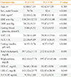
The subjects included 158 men (66.4%) and 80 women (33.6%), and the mean age was 42.8 years. Previously published studies suggested cutoff values for the FLI (i.e., FLI <30 as a rule-out criterion for fatty liver) [5]. Our subjects were divided into two groups according to FLI in 2007 using a cutoff score of 30, with 152 subjects <30 and 86 subjects ≥30. The baseline characteristics of the subjects who were subsequently diagnosed with NAFLD by FLI ≥30 and the characteristics of subjects with FLI <30 are shown in Table 1. As shown in Table 1, the mean ages of the group with FLI <30 (suggestive of non-NAFLD, i.e., normal) and the group with FLI ≥30 (suggestive of NAFLD) were not significantly different in 2003; however, the group with FLI ≥30 included a significantly higher proportion of male subjects (P<0.001).
The parameters indicating metabolic syndrome such as systolic blood pressure, diastolic blood pressure, fasting blood glucose, and triglycerides at baseline (in 2003) were significantly higher in the group with FLI ≥30 than the group with FLI <30 (P<0.001). In contrast, HDL-C levels were significantly lower in the group with FLI ≥30 (P<0.001). In addition, LDL-C, total cholesterol, fasting insulin, AST level, and ALT level were significantly higher in the group with FLI ≥30 (Table 1).
We also investigated the correlations between FLI at the 4-year follow-up and various baseline parameters, including A-FABP. In an unadjusted model, all parameters of metabolic markers had significant correlations with FLI. A-FABP also had a positive correlation with FLI. The baseline serum A-FABP was significantly associated with FLI after adjusting for age and sex (P<0.001). After adjusting for age and sex, we found that AST, ALT, fasting insulin, total cholesterol, and HDL-C were significantly correlated with FLI (Table 2). The association between FLI and ultrasonographic findings of NAFLD was significant. The median value of FLI for NAFLD on ultrasonography was higher than the normal finding on ultrasonography (26.05 [11.24 to 50.21] and 14.64 [4.79 to 37.61] respectively; P=0.006; values expressed as median [25th to 75th percentiles]).
When subjects were grouped into A-FABP tertiles, rising tertiles of A-FABP had significantly higher values of FLI even after adjusting for age and sex (P for trend <0.001) (Fig. 1). In the multiple logistic regression analysis with development of NAFLD defined by FLI greater than or equal to 30 as the dependent variable, the highest tertile of A-FABP had an increased likelihood of FLI ≥30 compared with the lowest tertile (odds ratio [OR], 1.97; P<0.05). After adjusting for age and sex, the OR for the risk of FLI ≥30 in the highest tertile of A-FABP maintained significance (OR, 2.68; P=0.012) (Table 3).

DISCUSSION

In this study, we found that serum A-FABP levels in healthy subjects are significantly associated with FLI and that there is a trend of increasing FLI score in proportion to serum A-FABP tertile. Subjects in the highest tertile of A-FABP were approximately three times more likely to have NAFLD compared with those in the lowest tertile in multiple logistic regression analysis, with development of NAFLD defined by FLI greater than or equal to 30.
Many studies have reported that A-FABP binds fatty acid ligands and functions in the regulation of lipid metabolism and gene expression [11-13]. Cao and colleagues [14] demonstrated that ob/ob-aP2-mal1-deficient mice were protected against fatty liver disease. Another study found that fatty infiltration of the liver was attenuated and total liver triglyceride content was reduced in aP2-inhibitor treated ob/ob mice [15]. In humans, there was one report about an association between A-FABP and fatty liver disease. Milner et al. [4] reported that increased serum A-FABP levels were a predictive factor for intrahepatic inflammation and fibrosis in patients with histologically confirmed NAFLD with abnormal liver function. They also reported an association between high serum A-FABP levels and progression of the disease, which was consistent with our results showing an association between A-FABP and FLI as a noninvasive marker of fatty liver disease.
Liver biopsy is the definitive method for diagnosing NAFLD, but this technique is an invasive procedure and is not completely precise due to sampling variability. Thus, some proxy markers such as the ratio of liver enzymes or ultrasonography are preferably used in epidemiology [16]. The FLI uses an equation with GGT, triglycerides, BMI, and waist circumference. It varies between 0 and 100 and is known to have significant accuracy in detecting NAFLD [17,18]. In addition, according to a study by Bedogni et al. [5], an FLI <30 rules out fatty liver. In this study, we divided the study subjects into two groups using an FLI of 30 as the cutoff value.
There are several limitations to the current study. First, the subjects in this study were selected from participants in a health check-up and are not representative of the general population. Second, there was a relatively large proportion of male subjects (66.4%). Third, we did not consider lifestyle risk factors such as exercise habits or diet. Despite with these limitations, we demonstrated a significant relationship between A-FABP levels and the development of NAFLD in healthy subjects at 4-year follow-up using the noninvasive marker FLI. Many studies on the association of A-FABP with type 2 diabetes or NAFLD as seen on ultrasonography have been reported; however, there is no study about the association between serum A-FABP levels and FLI in healthy Korean subjects.
In conclusion, this study shows that higher serum A-FABP levels at baseline were associated with FLI as a predictive indicator of NAFLD. In other words, high A-FABP levels were associated with a future risk of developing NAFLD. Further studies are needed to confirm the role of FLI as a predictive marker for NAFLD and its association with A-FABP.

Figures and Tables

Fig. 1
Association between adipocyte-specific fatty acid binding protein (A-FABP) tertile and fatty liver index (FLI) after adjusting for age and sex. Values are expressed as mean±SEM.
enm-28-283-g001
Table 1
Baseline Characteristics of Subjects in 2003 Grouped by Fatty Liver Index
enm-28-283-i001

Values are expressed as mean±SD.

FLI, fatty liver index; BMI, body mass index; SBP, systolic blood pressure; DBP, diastolic blood pressure; AST, aspartate aminotransferase; ALT, alanine aminotransferase; HDL-C, high density lipoprotein cholesterol; LDL-C, low density lipoprotein cholesterol; A-FABP, adipocyte-specific fatty acid binding protein.

Table 2
Correlation between Fatty Liver Index and Different Parameters
enm-28-283-i002

SBP, systolic blood pressure; DBP, diastolic blood pressure; AST, aspartate aminotransferase; ALT, alanine aminotransferase; HDL-C, high density lipoprotein cholesterol; LDL-C, low density lipoprotein cholesterol; A-FABP, adipocyte-specific fatty acid binding protein.

aModel 1, unadjusted; bModel 2, adjusted for age and sex.

Table 3
Multivariable Logistic Regression Analysis Showing Odds Ratios of Developing Nonalcoholic Fatty Liver Disease (Fatty Liver Index ≥30)
enm-28-283-i003

A-FABP, adipocyte-specific fatty acid binding protein; OR, odds ratio; CI, confidence interval.

aModel 1, unadjusted; bModel 2, adjusted for age and sex.

Notes

No potential conflict of interest relevant to this article was reported.

References

1. Tso AW, Xu A, Sham PC, Wat NM, Wang Y, Fong CH, Cheung BM, Janus ED, Lam KS. Serum adipocyte fatty acid binding protein as a new biomarker predicting the development of type 2 diabetes: a 10-year prospective study in a Chinese cohort. Diabetes Care. 2007; 30:2667–2672.
2. Terra X, Quintero Y, Auguet T, Porras JA, Hernandez M, Sabench F, Aguilar C, Luna AM, Del Castillo D, Richart C. FABP 4 is associated with inflammatory markers and metabolic syndrome in morbidly obese women. Eur J Endocrinol. 2011; 164:539–547.
3. Miyoshi T, Onoue G, Hirohata A, Hirohata S, Usui S, Hina K, Kawamura H, Doi M, Kusano KF, Kusachi S, Ninomiya Y. Serum adipocyte fatty acid-binding protein is independently associated with coronary atherosclerotic burden measured by intravascular ultrasound. Atherosclerosis. 2010; 211:164–169.
4. Milner KL, van der Poorten D, Xu A, Bugianesi E, Kench JG, Lam KS, Chisholm DJ, George J. Adipocyte fatty acid binding protein levels relate to inflammation and fibrosis in nonalcoholic fatty liver disease. Hepatology. 2009; 49:1926–1934.
5. Bedogni G, Bellentani S, Miglioli L, Masutti F, Passalacqua M, Castiglione A, Tiribelli C. The fatty liver index: a simple and accurate predictor of hepatic steatosis in the general population. BMC Gastroenterol. 2006; 6:33.
6. Musso G, Gambino R, Durazzo M, Cassader M. Noninvasive assessment of liver disease severity with liver fat score and CK-18 in NAFLD: prognostic value of liver fat equation goes beyond hepatic fat estimation. Hepatology. 2010; 51:715–717.
7. Kotronen A, Peltonen M, Hakkarainen A, Sevastianova K, Bergholm R, Johansson LM, Lundbom N, Rissanen A, Ridderstråle M, Groop L, Orho-Melander M, Yki-Jarvinen H. Prediction of non-alcoholic fatty liver disease and liver fat using metabolic and genetic factors. Gastroenterology. 2009; 137:865–872.
8. Bozkurt L, Gobl CS, Tura A, Chmelik M, Prikoszovich T, Kosi L, Wagner O, Roden M, Pacini G, Gastaldelli A, Kautzky-Willer A. Fatty liver index predicts further metabolic deteriorations in women with previous gestational diabetes. PLoS One. 2012; 7:e32710.
9. Osawa H, Mori Y. Sonographic diagnosis of fatty liver using a histogram technique that compares liver and renal cortical echo amplitudes. J Clin Ultrasound. 1996; 24:25–29.
10. Saadeh S, Younossi ZM, Remer EM, Gramlich T, Ong JP, Hurley M, Mullen KD, Cooper JN, Sheridan MJ. The utility of radiological imaging in nonalcoholic fatty liver disease. Gastroenterology. 2002; 123:745–750.
11. Coe NR, Bernlohr DA. Physiological properties and functions of intracellular fatty acid-binding proteins. Biochim Biophys Acta. 1998; 1391:287–306.
12. Chmurzynska A. The multigene family of fatty acid-binding proteins (FABPs): function, structure and polymorphism. J Appl Genet. 2006; 47:39–48.
13. Hertzel AV, Bernlohr DA. The mammalian fatty acid-binding protein multigene family: molecular and genetic insights into function. Trends Endocrinol Metab. 2000; 11:175–180.
14. Cao H, Maeda K, Gorgun CZ, Kim HJ, Park SY, Shulman GI, Kim JK, Hotamisligil GS. Regulation of metabolic responses by adipocyte/macrophage fatty acid-binding proteins in leptin-deficient mice. Diabetes. 2006; 55:1915–1922.
15. Furuhashi M, Tuncman G, Gorgun CZ, Makowski L, Atsumi G, Vaillancourt E, Kono K, Babaev VR, Fazio S, Linton MF, Sulsky R, Robl JA, Parker RA, Hotamisligil GS. Treatment of diabetes and atherosclerosis by inhibiting fatty-acid-binding protein aP2. Nature. 2007; 447:959–965.
16. Targher G, Day CP, Bonora E. Risk of cardiovascular disease in patients with nonalcoholic fatty liver disease. N Engl J Med. 2010; 363:1341–1350.
17. Balkau B, Lange C, Vol S, Fumeron F, Bonnet F. Group Study D.E.S.I.R. Nine-year incident diabetes is predicted by fatty liver indices: the French D.E.S.I.R. study. BMC Gastroenterol. 2010; 10:56.
18. Lerchbaum E, Gruber HJ, Schwetz V, Giuliani A, Moller R, Pieber TR, Obermayer-Pietsch B. Fatty liver index in polycystic ovary syndrome. Eur J Endocrinol. 2011; 165:935–943.
TOOLS
Similar articles