Journal List > Endocrinol Metab > v.27(3) > 1085951

Santoro, Carlomagno, and Melillo: RET: A Multi-Faceted Gene in Human Cancer

Abstract

REarranged during Transfection (RET) gene encodes a receptor tyrosine kinase and it was initially discovered as an in vitro transforming gene. For many years, RET has been involved in papillary thyroid carcinoma and medullary thyroid carcinoma. More recently, lung adenocarcinoma and chronic myelomonocytic leukemia samples have been found to display RET gene rearrangements. This knowledge is stimulating the search for protein kinase inhibitors to combat RET-driven malignancies.

REARRANGED DURING TRANSFECTION (RET) RECEPTOR AT A GLANCE

Receptor tyrosine kinases (RTK) are transmembrane (TM) proteins featuring an intracellular domain containing the tyrosine kinase (TK) enzyme. RTKs are often involved in cancer formation [1-3]. Notable examples are epidermal growth factor receptor (EGFR/HER1) and anaplastic lymphoma kinase (ALK) in non-small cell lung carcinoma (NSCLC) [4], KIT in gastrointestinal stromal tumors (GIST) [5], FLT3 in acute myeloid leukemia (AML) [6], and HER2/ERBB2/neu in breast cancer [7].
In some cases, cancer cells up-regulate expression of the RTK (as an example HER2 in breast cancer), its cognate growth factor or both, in other cases, structural alterations such as chromosomal rearrangements leading to the RTK recombination to heterologous genes (as an example EML4-ALK in lung adenocarcinoma) or point mutations (as EGFR, KIT or FLT3 mutations in NSCLC, GIST, or AML, respectively), lead to unchecked kinase and oncogenic activity [1-3].
This notion has stimulated the search for agents, such as monoclonal antibodies against the RTK extracellular domain (like trastuzumab for HER2 or cetuximab for EGFR) or ATP-competitive small molecule protein kinase inhibitors (PKIs) (like gefitinib and erlotinib for EGFR or crizotinib for ALK), to combat cancers driven by oncogenic RTKs [1-3].
The RET RTK was originally identified as an oncogene activated by a rearrangement occurred in vitro during transfection of NIH3T3 cells with human lymphoma DNA [8]. RET protein belongs to a cell-surface complex able to bind glial-derived neurotrophic factor (GDNF) ligands (GDNF, neurturin, artemin, and persephin) in conjunction with co-receptors of the GDNF receptor α family, designated GFRα 1-4 [9]. Binding to the ligand-co-receptor complex leads to RET dimerization and kinase activation. RET expression is tightly regulated during development and in the adulthood is limited to specific tissues, including neural crest-derived cells. RET is essential for the development of the enteric neurvous system and kidney, and germline loss-of-function mutations in RET cause Hirschsprung disease (aganglionic megacolon) and congenital anomalies of the kidney or lower urinary tract [10,11].
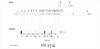
RET gene maps to chromosome 10q11.2. Fig. 1 shows that it is splitted in 21 coding exons. Exons 1-10 code for the extracellular region; exon 11 codes for the COOH-terminal part of the extracellular region, the TM domain, and the intracellular juxtamembrane domain. Finally, exons 12-21 code for the intracellular domain. An alternative splicing at exon 19 determine the synthesis of three RET protein isoforms with different C-terminal tails. In RET9 (1072 aa), exon 19 is unspliced; in RET51 (1114 aa), exon 19 is spliced to exon 20; in RET43 (1106 aa), exon 19 is spliced to to exon 21 [12-15]. RET9 and RET51 are the most abundant and well characterized isoforms (Fig. 1). RET protein features an extracellular portion (RET-EC) that includes the cleavable signal peptide (SP), four cadherin-like repeats (CLD1-4) and a cysteine-rich domain (CRD), a TM portion and an intracellular portion with the TK (RET-TK) domain split in two subdomains by a short insert (Fig. 1) [16,17].
The intracellular region of RET contains several tyrosine residues that undergo phosphorylation upon RET activation (Fig. 1) [18,19]. Tyrosine 905 map in the kinase A-loop (activation loop) and contributes to RET kinase activation; Y1015 is a docking site for phospholipase Cγ; Y1062 acts as a binding site for different proteins, including Shc, ShcC, IRS1/2, FRS2, and DOK1/4/5, that, in turn, lead to stimulation of the RAS/MAPK and phosphatidylinositol-3-kinase/AKT pathways [18-22].

RET POINT MUTATIONS IN CANCER

Activating point mutations of RET have been identified as a major driver for medullary thyroid carcinoma (MTC) [19,23-25]. MTC (about 5% of thyroid cancers) is a malignant tumor arising from neural crest derived thyroid parafollicular C cells. MTC occurs sporadically in 75% of the cases; in about 25% of cases, MTC is inherited as a component of the autosomal dominant multiple endocrine neoplasia type 2 (MEN2A, MEN2B, FMTC) syndromes [19,23-25]. MTC has full penetrance in all MEN subtypes and it is usually the first manifestation of the syndrome. MEN2A is the most common (80-90% of cases) subtype and is characterized by MTC, pheochromocytoma, and hyperparathyroidism. More rarely, MEN2A patients develop aganglionic megacolon (Hirschsprung disease) or cutaneous lichen [19]. MEN2B is the least common (5-10% of the cases) MEN2 subtype and it is characterized by aggressive MTC, pheochromocytoma, ganglioneuromatosis of the intestine, thickening of corneal nerves and marfanoid habitus. FMTC features MTC as the only phenotype and it is currently regarded as a low penetrance MEN2A subtype [24,25].
Germline RET mutations are responsible for virtually all MEN2 cases. Most common mutations target exons 10 and 11 encoding CRD of the RET extracellular domain or exons 13-16 encoding part of the TK domain of RET (Fig. 1). Most frequent (85% of the cases) MEN2A mutations affect cysteine 634 (in exon 11) in the CRD; less commonly, MEN2A is caused by mutations of cysteines C609, C611, C618, C620 (in exon 10), or C630 (in exon 11) [19]. Other rare single or double mutations, small insertions or deletions, have been described in MEN2A cases [19]. FMTC mutations are evenly distributed among the various cysteines of CRD [19]. FMTC can be also associated to mutations of the RET-TK (E768D, L790F, Y791F, V804L, V804M, and S891A) (Fig. 1). MEN2B mutation is caused in most (> 97%) of the cases by M918T mutation in RET-TK (exon 16); more rarely (2%), MEN2B patients harbor the A883F substitution (exon 15), or double mutations (Fig. 1) [19]. Importantly, RET mutations (mainly M918T) occur at the somatic level in about half sporadic MTC.
MTC-associated RET mutations have a gain-of-function effect and convert RET into an oncogene. Extracellular cysteine RET mutants form covalent dimers stabilized by disulfide-bonds and display growth factor independent kinase activity [26]. In unstimulated conditions, the RET-TK adopts a trans-inhibited head-to-tail inactive dimer conformation in which the substrate-binding site of each monomer is occluded by the contralateral one [17]. Some mutations targeting the TK domain (most notably M918T) hit trans-inhibited dimer contact points and may therefore destabilize this inactive dimer conformation and activate RET [17]. Moreover, methionine 918 localizes in the P + 1 kinase loop, a site that is involved in substrate binding. Accordingly, its replacement by a threonine residue modifies RET signaling specificity [26,27].

RET REARRANGEMENTS IN CANCER

At least three types of human cancer (papillary thyroid carcinoma [PTC], lung adenocarcinoma, and chronic myelomonocytic leukemia [CMML]) feature genomic rearrangements leading to the recombination of the RET-TK domain to heterologous proteins. Breakpoint in RET is virtually always in intron 11, so that RET exon 12 (encoding the N-ter of the RET-TK) is fused to the 5'-end of heterologous genes (Fig. 1). Fusion to heterologous proteins containing protein homodimerization motives results in constitutive RET kinase dimerization, growth factor independent activation, and signalling. Furthermore, replacement of the RET transcriptional promoter with those of the RET fusion partners likely de-regulates RET expression. The expression of a constitutively dimerized and active RET kinase leads to chronic exposure of cancer cells to the activation of intracellular signalling pathways, such as the RAS-RAF-MAPK, that are activated by RET [18]. Intriguingly, this pathway includes RAS and BRAF that are very commonly mutated in the same cancer types in which RET is involved [4,28].
RET gene rearrangements were initially discovered in PTC [29]. PTC arises from follicular thyroid cells and is the most prevalent thyroid cancer type [28]. In PTC, chromosomal aberrations, most commonly a paracentric inversion of the long arm of chromosome 10, cause the illegitimate recombination of the RET-TK (from exon 12 to the 3'-end) to the promoter sequence and 5'-terminal exons of heterologous genes [28]. Most common RET/PTC rearrangements (90% of the cases) are RET/PTC1 (CCDC6-RET) and RET/PTC3 (NCOA4-RET) [30,31]. RET/PTC3 is particularly frequent in PTC consequent to the Chernobyl disaster and in young patients [28]. Close proximity of the fusion partners in thyrocyte chromatin may favour their recombination [32,33]. RET/PTC prevalence (average 25% of the cases) varies considerably in different patient series [28]. An important factor for this variability is methodology used for the detection [34].
More recently, RET has been demonstrated to play an important role also in a subset of NSCLC cases, in particular in lung adenocarcinoma. In about 1% NSCLC, inversions of chromosome 10 cause the fusion of the RET-TK domain to different 5'-terminal exons (15, 16, 22, 23, or 24) of KIF5B (kinesin family member 5B) gene [35-37]. The RET/PTC1 (CCDC6-RET chimera) oncogene has been found in one lung adenocarcinoma sample [38]. As in the case of RET/PTC, also KIF5B-RET fusion proteins likely form active homodimers through the coiled-coil domain present in the NH2-ter portion of KIF5B. It is important to note that lung adenocarcinoma is commonly associated to mutations targeting also RTKs other than RET, such as EGFR, ROS1, and ALK [4]. Mutations in EGFR, ROS1, ALK, and RET are mutually exclusive.
CMML is a neoplastic myeloid disorder [39]. Very recently, gene rearrangements causing the fusion of the RET-encoding TK domain (from exon 12) in one case to the 5'-terminal four exons of break-point cluster region (BCR) and in another case to the 5'-terminal 12 exons of fibroblast growth factor receptor 1 oncogenic partner (FGFR1OP) genes have been described in CMML [40]. Prevalence of RET rearrangements in CMML is still unknown.

RET OVER-EXPRESSION IN CANCER

In some cancers, RET upregulation rather than structural alteration has been reported. This is worthmentioning in the case of breast and pancreatic adenocarcinoma. A positive correlation was demonstrated between RET over-expression and estrogen receptor-positive breast carcinoma [41,42]. Importantly, RET inhibition restored a hormone-sensitive phenotype in anti-estrogen resistant breast cancer cells [43]. Furthermore, RET protein was overexpressed in pancreatic carcinoma and involved in neural invasion of pancreatic cancer cells [44,45].

RET AS A TUMOR SUPPRESSOR

In contrast to its well-established role as an oncogene for several cancer types, RET has been recently proposed to play tumor suppressor roles in colorectal cancer (CRC) and pituitary adenoma [46, 47]. Such tumor suppressor role might be functionally linked to a pro-apoptotic role exerted by RET by behaving as a "dependence" receptor [48]. Dependence receptors display pro-apoptotic activity when not bound to cognate growth factor; in the case of RET, this leads to caspase-3-mediated cleavage of its cytosolic portion (after aspartic acid residues 707 and 1017) which, in turn, releases a cytosolic peptide (aa 708-1016) that is able to induce cell death [48]. Thus, loss-of-function of RET may abrogate this effect and foster tumor development.
In CRC, RET promoter methylation commonly silenced RET expression [47]. Moreover, in rare CRC samples, somatic mutations (V145G, R360W, and G593E) in RET extracellular domain impaired RET-mediated apoptosis of colon epithelial cells; thus, either RET downregulation or mutations causing loss of RET-mediated apoptosis may be selected during CRC formation [47].
Similarly, RET was expressed in somatotroph-derived pituitary adenomas, where it acted as a two-sided tumor regulator. When stimulated by GDNF, it behaved as as an oncogene able to activate intracellular signaling and cell survival. Instead, in the absence of GDNF, RET behaved as a tumor suppressor; caspase-mediated RET processing induced Pit-1 expression, that, in turn, caused p19Arf and p53 upregulation and apoptosis [46,49].

RET KINASE INHIBITORS FOR CANCER TREATMENT

The advent of small-molecule drugs and monoclonal antibodies made RTK targeting a feasible cancer therapeutic strategy [2]. Most RTK-directed small-molecule drugs are PKIs that obstruct kinase activity by binding to the ATP pocket of the kinase in competition with cellular ATP [2,3]. Prototypic examples of anti-neoplastic PKIs are imatinib, an inhibitor of ABL, KIT and platelet-derived growth factor receptor (PDGFR), in BCR-ABL-positive chronic myelogenous leukemia and KIT or PDGFR-α mutant GISTs and EGFR-directed inhibitors (gefitinib and erlotinib) for EGFR mutant lung adenocarcinoma [4,50].
Several small-molecules have been identified at the preclinical level to target cancer cells showing increased RET activity [51-53]. These agents are multitargeted and able to inhibit several kinases besides RET. Vandetanib (ZD6474) is an anilinoquinazoline that docks in the ATP-binding pocket of the RET kinase and inhibits RET kinase with an inhibitory concentration 50 of 100-130 nM [17, 54]. Some RET mutations, like V804M/L, and Y806C cause resistance to [55,56]. Other compounds with anti-RET activity inlcude sorafenib, sunitinib, lenvatinib (E7080), and cabozantinib (XL-184) [57-60]. Some of them already entered clinical experimentation [61-63]. These compounds share with vandetanib the capability of targeting vascular endothelial growth factor receptors (VEGFR2/KDR; VEGFR3/Flt-4, VEGFR1/Flt-1) [64,65]. In addition, vandetanib targets the EGFR [66]. Based on the results of the ZETA trial, vandetanib has been recently registered for locally advanced or metastatic MTC [62] and may represent a promising agent for other RET-driven cancers.
As most cancers are the result of a number of mutations and feature multiple altered signaling pathways, it may be anticipated that PKIs able to target multiple kinases or a rational combination of them will be more clinically effective than agents blocking a single kinase. Multi-targeting or combination therapies may also attenuate resistance formation [67]. By using a chemical genetic approach and a Drosophila model of MEN2 new PKIs able to inhibit simultaneously RET, RAF, SRC, and S6K have shown increased potency and reduced toxicity [68].

CONCLUSIONS

After about three decades since its discovery [8], RET has raised a great interest as a gene involved in human developmental diseases as well as epithelial, neuroendocrine and hematological cancers. This knowledge has been already transferred to the bed of patients, as illustrated by RET genotyping to identify MEN2 carriers [69]. Moreover, this knowledge has also led to the use of RET-directed therapeutics for the treatment of thyroid cancer.

Figures and Tables

Fig. 1
Schematic drawing of the RET gene (upper) and protein (lower). RET gene structure with coding exons is represented; nucleotide (nt) positions are marked. An alternative splicing in exon 19 generate two alternative mRNAs, coding for RET-51 (1,114 residues), when exon 19 is spliced to exon 20, or RET-9 (1,072 residues), when exon 19 remains unspliced. Last coding exon (exon 21) that encodes the C-terminal part of another less abundant RET isoform, RET (43), is not represented. Position of the STOP codons for RET-51 and RET-9 is marked. RET protein is represented in the bottom part of the figure. The extracellular RET domain (with the signal peptide [SP], four cadherin-like repeats [CLD1-4] and a cysteine-rich domain [CRD]), the transmembrane domain (TM), and the intracellular tyrosine kinase domain (TK) are represented. The RET-TK is split into two subdomains (TK1 and 2) by an insert region (KI). The position of most common RET point mutations associated to familial and sporadic MTC is shown. The breakpoint in RET (at the boundary between exons 11 and 12), that is involved in RET gene rearrangements in papillary thyroid carcinoma, lung adenocarcinoma, and chronic myelomonocytic leukemia, is represented. All these informations were retrieved from National Center for Biotechnology Information, with the exception of RET kinase insert boundaries that were retrieved from Knowles et al. [17] and cadherin-like domain boundaries that were retrieved from Scott et al. [16].
enm-27-173-g001

ACKNOWLEDGEMENTS

We gratefully acknowledge members of our laboratory for continuous support.

References

1. Krause DS, Van Etten RA. Tyrosine kinases as targets for cancer therapy. N Engl J Med. 2005. 353:172–187.
2. Baselga J. Targeting tyrosine kinases in cancer: the second wave. Science. 2006. 312:1175–1178.
3. Zhang J, Yang PL, Gray NS. Targeting cancer with small molecule kinase inhibitors. Nat Rev Cancer. 2009. 9:28–39.
4. Herbst RS, Heymach JV, Lippman SM. Lung cancer. N Engl J Med. 2008. 359:1367–1380.
5. Corless CL, Barnett CM, Heinrich MC. Gastrointestinal stromal tumours: origin and molecular oncology. Nat Rev Cancer. 2011. 11:865–878.
6. Daver N, Cortes J. Molecular targeted therapy in acute myeloid leukemia. Hematology. 2012. 17:Suppl 1. S59–S62.
7. Arribas J, Baselga J, Pedersen K, Parra-Palau JL. p95HER2 and breast cancer. Cancer Res. 2011. 71:1515–1519.
8. Takahashi M, Ritz J, Cooper GM. Activation of a novel human transforming gene, ret, by DNA rearrangement. Cell. 1985. 42:581–588.
9. Airaksinen MS, Saarma M. The GDNF family: signalling, biological functions and therapeutic value. Nat Rev Neurosci. 2002. 3:383–394.
10. Runeberg-Roos P, Saarma M. Neurotrophic factor receptor RET: structure, cell biology, and inherited diseases. Ann Med. 2007. 39:572–580.
11. Jain S. The many faces of RET dysfunction in kidney. Organogenesis. 2009. 5:177–190.
12. Tahira T, Shiraishi M, Ishizaka Y, Ikeda I, Sakai R, Sugimura T, Nagao M. A TaqI RFLP in the human ret proto-oncogene. Nucleic Acids Res. 1990. 18:7472.
13. Myers SM, Eng C, Ponder BA, Mulligan LM. Characterization of RET proto-oncogene 3' splicing variants and polyadenylation sites: a novel C-terminus for RET. Oncogene. 1995. 11:2039–2045.
14. Ivanchuk SM, Eng C, Cavenee WK, Mulligan LM. The expression of RET and its multiple splice forms in developing human kidney. Oncogene. 1997. 14:1811–1818.
15. Ivanchuk SM, Myers SM, Mulligan LM. Expression of RET 3' splicing variants during human kidney development. Oncogene. 1998. 16:991–996.
16. Scott RP, Ibanez CF. Determinants of ligand binding specificity in the glial cell line-derived neurotrophic factor family receptor alpha S. J Biol Chem. 2001. 276:1450–1458.
17. Knowles PP, Murray-Rust J, Kjaer S, Scott RP, Hanrahan S, Santoro M, Ibáñez CF, McDonald NQ. Structure and chemical inhibition of the RET tyrosine kinase domain. J Biol Chem. 2006. 281:33577–33587.
18. Takahashi M. The GDNF/RET signaling pathway and human diseases. Cytokine Growth Factor Rev. 2001. 12:361–373.
19. de Groot JW, Links TP, Plukker JT, Lips CJ, Hofstra RM. RET as a diagnostic and therapeutic target in sporadic and hereditary endocrine tumors. Endocr Rev. 2006. 27:535–560.
20. Melillo RM, Santoro M, Ong SH, Billaud M, Fusco A, Hadari YR, Schlessinger J, Lax I. Docking protein FRS2 links the protein tyrosine kinase RET and its oncogenic forms with the mitogen-activated protein kinase signaling cascade. Mol Cell Biol. 2001. 21:4177–4187.
21. Melillo RM, Carlomagno F, De Vita G, Formisano P, Vecchio G, Fusco A, Billaud M, Santoro M. The insulin receptor substrate (IRS)-1 recruits phosphatidylinositol 3-kinase to Ret: evidence for a competition between Shc and IRS-1 for the binding to Ret. Oncogene. 2001. 20:209–218.
22. Melillo RM, Castellone MD, Guarino V, De Falco V, Cirafici AM, Salvatore G, Caiazzo F, Basolo F, Giannini R, Kruhoffer M, Orntoft T, Fusco A, Santoro M. The RET/PTC-RAS-BRAF linear signaling cascade mediates the motile and mitogenic phenotype of thyroid cancer cells. J Clin Invest. 2005. 115:1068–1081.
23. Wells SA Jr, Santoro M. Targeting the RET pathway in thyroid cancer. Clin Cancer Res. 2009. 15:7119–7123.
24. Eng C. Common alleles of predisposition in endocrine neoplasia. Curr Opin Genet Dev. 2010. 20:251–256.
25. Eng C. Mendelian genetics of rare: and not so rare: cancers. Ann N Y Acad Sci. 2010. 1214:70–82.
26. Santoro M, Carlomagno F, Romano A, Bottaro DP, Dathan NA, Grieco M, Fusco A, Vecchio G, Matoskova B, Kraus MH, Di Fiore PP. Activation of RET as a dominant transforming gene by germline mutations of MEN2A and MEN2B. Science. 1995. 267:381–383.
27. Songyang Z, Carraway KL 3rd, Eck MJ, Harrison SC, Feldman RA, Mohammadi M, Schlessinger J, Hubbard SR, Smith DP, Eng C, Lorenzo MA, Ponder BA, Mayer BJ, Cantley LC. Catalytic specificity of protein-tyrosine kinases is critical for selective signalling. Nature. 1995. 373:536–539.
28. Nikiforov YE, Nikiforova MN. Molecular genetics and diagnosis of thyroid cancer. Nat Rev Endocrinol. 2011. 7:569–580.
29. Fusco A, Grieco M, Santoro M, Berlingieri MT, Pilotti S, Pierotti MA, Della Porta G, Vecchio G. A new oncogene in human thyroid papillary carcinomas and their lymph-nodal metastases. Nature. 1987. 328:170–172.
30. Grieco M, Santoro M, Berlingieri MT, Melillo RM, Donghi R, Bongarzone I, Pierotti MA, Della Porta G, Fusco A, Vecchio G. PTC is a novel rearranged form of the ret proto-oncogene and is frequently detected in vivo in human thyroid papillary carcinomas. Cell. 1990. 60:557–563.
31. Santoro M, Dathan NA, Berlingieri MT, Bongarzone I, Paulin C, Grieco M, Pierotti MA, Vecchio G, Fusco A. Molecular characterization of RET/PTC3; a novel rearranged version of the RETproto-oncogene in a human thyroid papillary carcinoma. Oncogene. 1994. 9:509–516.
32. Nikiforova MN, Stringer JR, Blough R, Medvedovic M, Fagin JA, Nikiforov YE. Proximity of chromosomal loci that participate in radiation-induced rearrangements in human cells. Science. 2000. 290:138–141.
33. Gandhi M, Evdokimova VN, K TC, Nikiforova MN, Kelly LM, Stringer JR, Bakkenist CJ, Nikiforov YE. Homologous chromosomes make contact at the sites of double-strand breaks in genes in somatic G0/G1-phase human cells. Proc Natl Acad Sci U S A. 2012. 109:9454–9459.
34. Zhu Z, Ciampi R, Nikiforova MN, Gandhi M, Nikiforov YE. Prevalence of RET/PTC rearrangements in thyroid papillary carcinomas: effects of the detection methods and genetic heterogeneity. J Clin Endocrinol Metab. 2006. 91:3603–3610.
35. Lipson D, Capelletti M, Yelensky R, Otto G, Parker A, Jarosz M, Curran JA, Balasubramanian S, Bloom T, Brennan KW, Donahue A, Downing SR, Frampton GM, Garcia L, Juhn F, Mitchell KC, White E, White J, Zwirko Z, Peretz T, Nechushtan H, Soussan-Gutman L, Kim J, Sasaki H, Kim HR, Park SI, Ercan D, Sheehan CE, Ross JS, Cronin MT, Jänne PA, Stephens PJ. Identification of new ALK and RET gene fusions from colorectal and lung cancer biopsies. Nat Med. 2012. 18:382–384.
36. Takeuchi K, Soda M, Togashi Y, Suzuki R, Sakata S, Hatano S, Asaka R, Hamanaka W, Ninomiya H, Uehara H, Lim Choi Y, Satoh Y, Okumura S, Nakagawa K, Mano H, Ishikawa Y. RET, ROS1 and ALK fusions in lung cancer. Nat Med. 2012. 18:378–381.
37. Kohno T, Ichikawa H, Totoki Y, Yasuda K, Hiramoto M, Nammo T, Sakamoto H, Tsuta K, Furuta K, Shimada Y, Iwakawa R, Ogiwara H, Oike T, Enari M, Schetter AJ, Okayama H, Haugen A, Skaug V, Chiku S, Yamanaka I, Arai Y, Watanabe S, Sekine I, Ogawa S, Harris CC, Tsuda H, Yoshida T, Yokota J, Shibata T. KIF5B-RET fusions in lung adenocarcinoma. Nat Med. 2012. 18:375–377.
38. Li F, Feng Y, Fang R, Fang Z, Xia J, Han X, Liu XY, Chen H, Liu H, Ji H. Identification of RET gene fusion by exon array analyses in "pan-negative" lung cancer from never smokers. Cell Res. 2012. 22:928–931.
39. Vardiman JW, Thiele J, Arber DA, Brunning RD, Borowitz MJ, Porwit A, Harris NL, Le Beau MM, Hellström-Lindberg E, Tefferi A, Bloomfield CD. The 2008 revision of the World Health Organization (WHO) classification of myeloid neoplasms and acute leukemia: rationale and important changes. Blood. 2009. 114:937–951.
40. Ballerini P, Struski S, Cresson C, Prade N, Toujani S, Deswarte C, Dobbelstein S, Petit A, Lapillonne H, Gautier EF, Demur C, Lippert E, Pages P, Mansat-De Mas V, Donadieu J, Huguet F, Dastugue N, Broccardo C, Perot C, Delabesse E. RET fusion genes are associated with chronic myelomonocytic leukemia and enhance monocytic differentiation. Leukemia. In press 2012.
41. Tozlu S, Girault I, Vacher S, Vendrell J, Andrieu C, Spyratos F, Cohen P, Lidereau R, Bieche I. Identification of novel genes that co-cluster with estrogen receptor alpha in breast tumor biopsy specimens, using a large-scale real-time reverse transcription-PCR approach. Endocr Relat Cancer. 2006. 13:1109–1120.
42. Esseghir S, Todd SK, Hunt T, Poulsom R, Plaza-Menacho I, Reis-Filho JS, Isacke CM. A role for glial cell derived neurotrophic factor induced expression by inflammatory cytokines and RET/GFR alpha 1 receptor up-regulation in breast cancer. Cancer Res. 2007. 67:11732–11741.
43. Plaza-Menacho I, Morandi A, Robertson D, Pancholi S, Drury S, Dowsett M, Martin LA, Isacke CM. Targeting the receptor tyrosine kinase RET sensitizes breast cancer cells to tamoxifen treatment and reveals a role for RET in endocrine resistance. Oncogene. 2010. 29:4648–4657.
44. Veit C, Genze F, Menke A, Hoeffert S, Gress TM, Gierschik P, Giehl K. Activation of phosphatidylinositol 3-kinase and extracellular signal-regulated kinase is required for glial cell line-derived neurotrophic factor-induced migration and invasion of pancreatic carcinoma cells. Cancer Res. 2004. 64:5291–5300.
45. Gil Z, Cavel O, Kelly K, Brader P, Rein A, Gao SP, Carlson DL, Shah JP, Fong Y, Wong RJ. Paracrine regulation of pancreatic cancer cell invasion by peripheral nerves. J Natl Cancer Inst. 2010. 102:107–118.
46. Cañibano C, Rodriguez NL, Saez C, Tovar S, Garcia-Lavandeira M, Borrello MG, Vidal A, Costantini F, Japon M, Dieguez C, Alvarez CV. The dependence receptor Ret induces apoptosis in somatotrophs through a Pit-1/p53 pathway, preventing tumor growth. EMBO J. 2007. 26:2015–2028.
47. Luo Y, Tsuchiya KD, Il Park D, Fausel R, Kanngurn S, Welcsh P, Dzieciatkowski S, Wang J, Grady WM. RET is a potential tumor suppressor gene in colorectal cancer. Oncogene. In press 2012.
48. Bordeaux MC, Forcet C, Granger L, Corset V, Bidaud C, Billaud M, Bredesen DE, Edery P, Mehlen P. The RET proto-oncogene induces apoptosis: a novel mechanism for Hirschsprung disease. EMBO J. 2000. 19:4056–4063.
49. Diaz-Rodriguez E, García-Lavandeira M, Perez-Romero S, Senra A, Cañibano C, Palmero I, Borrello MG, Dieguez C, Alvarez CV. Direct promoter induction of p19Arf by Pit-1 explains the dependence receptor RET/Pit-1/p53-induced apoptosis in the pituitary somatotroph cells. Oncogene. 2012. 31:2824–2835.
50. Druker B. Imatinib (Gleevec) as a paradigm of targeted cancer therapies. Keio J Med. 2010. 59:1–3.
51. Phay JE, Shah MH. Targeting RET receptor tyrosine kinase activation in cancer. Clin Cancer Res. 2010. 16:5936–5941.
52. Sherman SI. Targeted therapies for thyroid tumors. Mod Pathol. 2011. 24:Suppl 2. S44–S52.
53. Gild ML, Bullock M, Robinson BG, Clifton-Bligh R. Multikinase inhibitors: a new option for the treatment of thyroid cancer. Nat Rev Endocrinol. 2011. 7:617–624.
54. Carlomagno F, Vitagliano D, Guida T, Ciardiello F, Tortora G, Vecchio G, Ryan AJ, Fontanini G, Fusco A, Santoro M. ZD6474, an orally available inhibitor of KDR tyrosine kinase activity, efficiently blocks oncogenic RET kinases. Cancer Res. 2002. 62:7284–7290.
55. Carlomagno F, Guida T, Anaganti S, Vecchio G, Fusco A, Ryan AJ, Billaud M, Santoro M. Disease associated mutations at valine 804 in the RET receptor tyrosine kinase confer resistance to selective kinase inhibitors. Oncogene. 2004. 23:6056–6063.
56. Carlomagno F, Guida T, Anaganti S, Provitera L, Kjaer S, McDonald NQ, Ryan AJ, Santoro M. Identification of tyrosine 806 as a molecular determinant of RET kinase sensitivity to ZD6474. Endocr Relat Cancer. 2009. 16:233–241.
57. Carlomagno F, Anaganti S, Guida T, Salvatore G, Troncone G, Wilhelm SM, Santoro M. BAY 43-9006 inhibition of oncogenic RET mutants. J Natl Cancer Inst. 2006. 98:326–334.
58. Kim DW, Jo YS, Jung HS, Chung HK, Song JH, Park KC, Park SH, Hwang JH, Rha SY, Kweon GR, Lee SJ, Jo KW, Shong M. An orally administered multitarget tyrosine kinase inhibitor, SU11248, is a novel potent inhibitor of thyroid oncogenic RET/papillary thyroid cancer kinases. J Clin Endocrinol Metab. 2006. 91:4070–4076.
59. Verbeek HH, Alves MM, de Groot JW, Osinga J, Plukker JT, Links TP, Hofstra RM. The effects of four different tyrosine kinase inhibitors on medullary and papillary thyroid cancer cells. J Clin Endocrinol Metab. 2011. 96:E991–E995.
60. Koh YW, Shah MH, Agarwal K, McCarty SK, Koo BS, Brendel VJ, Wang C, Porter K, Jarjoura D, Saji M, Ringel MD. Sorafenib and Mek inhibition is synergistic in medullary thyroid carcinoma in vitro. Endocr Relat Cancer. 2012. 19:29–38.
61. Wells SA Jr, Gosnell JE, Gagel RF, Moley J, Pfister D, Sosa JA, Skinner M, Krebs A, Vasselli J, Schlumberger M. Vandetanib for the treatment of patients with locally advanced or metastatic hereditary medullary thyroid cancer. J Clin Oncol. 2010. 28:767–772.
62. Wells SA Jr, Robinson BG, Gagel RF, Dralle H, Fagin JA, Santoro M, Baudin E, Elisei R, Jarzab B, Vasselli JR, Read J, Langmuir P, Ryan AJ, Schlumberger MJ. Vandetanib in patients with locally advanced or metastatic medullary thyroid cancer: a randomized, double-blind phase III trial. J Clin Oncol. 2012. 30:134–141.
63. Leboulleux S, Bastholt L, Krause T, de la Fouchardiere C, Tennvall J, Awada A, Gomez JM, Bonichon F, Leenhardt L, Soufflet C, Licour M, Schlumberger MJ. Vandetanib in locally advanced or metastatic differentiated thyroid cancer: a randomised, double-blind, phase 2 trial. Lancet Oncol. 2012. 13:897–905.
64. Lam ET, Ringel MD, Kloos RT, Prior TW, Knopp MV, Liang J, Sammet S, Hall NC, Wakely PE Jr, Vasko VV, Saji M, Snyder PJ, Wei L, Arbogast D, Collamore M, Wright JJ, Moley JF, Villalona-Calero MA, Shah MH. Phase II clinical trial of sorafenib in metastatic medullary thyroid cancer. J Clin Oncol. 2010. 28:2323–2330.
65. Kurzrock R, Sherman SI, Ball DW, Forastiere AA, Cohen RB, Mehra R, Pfister DG, Cohen EE, Janisch L, Nauling F, Hong DS, Ng CS, Ye L, Gagel RF, Frye J, Muller T, Ratain MJ, Salgia R. Activity of XL184 (Cabozantinib), an oral tyrosine kinase inhibitor, in patients with medullary thyroid cancer. J Clin Oncol. 2011. 29:2660–2666.
66. Wedge SR, Ogilvie DJ, Dukes M, Kendrew J, Chester R, Jackson JA, Boffey SJ, Valentine PJ, Curwen JO, Musgrove HL, Graham GA, Hughes GD, Thomas AP, Stokes ES, Curry B, Richmond GH, Wadsworth PF, Bigley AL, Hennequin LF. ZD6474 inhibits vascular endothelial growth factor signaling, angiogenesis, and tumor growth following oral administration. Cancer Res. 2002. 62:4645–4655.
67. Knight ZA, Lin H, Shokat KM. Targeting the cancer kinome through polypharmacology. Nat Rev Cancer. 2010. 10:130–137.
68. Dar AC, Das TK, Shokat KM, Cagan RL. Chemical genetic discovery of targets and anti-targets for cancer polypharmacology. Nature. 2012. 486:80–84.
69. American Thyroid Association Guidelines Task Force. Kloos RT, Eng C, Evans DB, Francis GL, Gagel RF, Gharib H, Moley JF, Pacini F, Ringel MD, Schlumberger M, Wells SA Jr. Medullary thyroid cancer: management guidelines of the American Thyroid Association. Thyroid. 2009. 19:565–612.
TOOLS
Similar articles