

 PDF
PDF ePub
ePub Citation
 Print
Print


INTRODUCTION
Due to recent progress in the abdominal imaging techniques and its widespread use, clinicians often encounter incidentally discovered adrenal mass [1,2]. Although there are wide variations in its prevalence depending on the study population and diagnosing modality, it is generally known to be about 4-6% [3-5]. The majority of adrenal incidentalomas are benign non-functioning adenoma. From the radiologic point of view, cystic lesions are rare constituting about 1-1.9% of adrenal incidentalomas [4,6,7].
We experienced a patient presenting with cystic adrenal incidentaloma, which finally turned out to be paragonimiasis. This is an extremely rare cause of adrenal infection and therefore we report it with a review of relevant literatures.
CASE REPORT
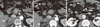
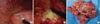
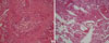
A 52-year-old male presented with fatigue and a weight loss of 5 kg over 6 months. The patient was referred for the evaluation of adrenal incidentaloma detected by ultrasonography; other malignancy check up did not show abnormality. He had past histories of type 2 diabetes and hypertension which had been treated with oral hypoglycemic agents and an antihypertensive agent for the previous 4 years. There were neither histories of other endocrine diseases nor infectious diseases. He did not drink alcohol. On admission, his vital signs were stable without fever. No symptoms and signs suggested Cushing's syndrome or pheochromocytoma. Laboratory findings revealed no abnormalities, except leukocytosis (white blood count 10,130/cm), eosinophilia (8.5%) and increased absolute eosinophil count (910/µL). In the evaluation of the functionality of adrenal mass, Cushing's syndrome was excluded by the tests for 24-hour urine free cortisol (55.9 µg/day) and 1 mg overnight dexamethasone suppression test (< 1.0 µg/dL). Primary aldosteronism was also excluded by PAC/PRA ratio (0.63). Concentrations of vanillylmandelic acid (2.87 mg/day), metanephrine (0.25 mg/day) from 24-hour urine collection excluded the possibility of pheochromocytoma. Chest X-ray was normal but computed tomography (CT) scan showed 6.5 × 5 cm sized multilocular septated cystic mass in right adrenal gland (Fig. 1A, B). Multiple, variable sized cystic lesions were also noted in the gastrocolic ligament, transverse mesocolon, and upper omentum areas (Fig. 1B, C). Although there was no hormonal overproduction, laparoscopic adrenalectomy was done due to its large size and for the confirmatory diagnosis. In the surgical field, multilobulated cystic adrenal mass was observed above the upper pole of right kidney and whitish patches were spread out in peritoneum, omentum, dome of liver and diaphragm (Fig. 2A). Right adrenal gland was totally replaced by cystic mass filled with mucopurulent creamy materials (Fig. 2B). Cross section of surgical specimen revealed variable-sized, multicystic mass (Fig. 2C). Light microscopic examination revealed chronic granulomatous inflammation with central necrosis containing numerous eggs of Paragonimus spp (Fig. 3). The eggs of Paragonimus spp were also found in peritoneum specimen. For immunologic diagnosis, ELISA was performed and the IgG titer of paragonimus antibody was positive (8.90) (Table 1).
Retrospective history taking revealed that he had ingested a raw crabs-preserved in soy sauce in Seomjin river, 2 years ago. His wife, eaten together had visited another hospital a year ago due to right-side pleural effusion and diagnosed as P. westermani infection.
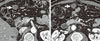
He was treated with praziquantel at a total dose of 150 mg per kg of body weight divided into three doses per day for 2 days. Follow-up CT scan taken 5 months (Fig. 4) after medical treatment showed little interval change of cystic lesions in the abdominal cavity. However, the proportion of eosinophils (4.7%), eosinophil count (360/µL) and IgG titer of paragonimus antibody (2.28) were significantly decreased (Table 1).
DISCUSSION
We report here a rare case of adrenal infection, paragonimiasis. To our knowledge, there have been only 2 cases described in the literature [8,9]. In our case, the diagnosis of paragonimiasis was supported not only by pathologic findings but also by serologic testing and the history of raw crab ingestion in the endemic area. This is an unusual finding in where pulmonary parasitism was not detected.
Although adrenal incidentaloma is frequently found in the daily practice, diagnosed in up to 4-6% of examinees [3-5], cystic lesions are rarely found. In one series of 1049 incidental adrenal lesions detected by CT scan, only 13 (1%) lesions were cystic [4,6]. The causes of adrenal cysts include lymphangioma, hemangioma, cystic adenoma and infections.
While various microbial pathogens can be infected, parasitic infection of the adrenal gland rarely occurs. They can involve multiple organs concomitantly, but isolated adrenal gland infection may take place. Not only immunocompromised patients but also immunocompetent hosts can be infested. There are several case reports of hydatid disease caused by Echinococcus spp, and it is estimated to account for 6-7% of adrenal cysts [10-12]. Other causes include Leishmania spp, Trypanosoma spp, Microsporidia spp and amebic species which also can present as a cystic form [7]. However, the incidence of the adrenal paragonimiasis is extremely rare. Basically, cystic lesions caused by Paragonimus could not be resolved by drug treatment. Rather, shrinkage of mass size might be resultant from healing process with residual scar formation [13]. Treatment of adrenal paragonimiasis and pulmonary paragonimiasis is not different at all. Due to lack of cases, however, nothing much is known about the progress and prognosis of adrenal paragonimiasis, except that cystic lesion may remain in abdominal cavity.
Paragonimiasis is endemic in African and Asian countries such as China, Japan, Taiwan and Korea. The nationwide prevalence of human paragonimiasis in China was estimated to be 1.71% [14]. It is also known to be widely distributed in Korea, although the prevalence is gradually decreasing [15]. With the possibility of developing extrapulmonary paragonimiasis, involvement of brain, spinal cord, subcutaneous tissue and urinary tract have been reported in Korea [15]. However, as the infestation of organs such as omentum and adrenal gland are less likely to produce definite symptoms, diagnosis of abdominal paragonimiasis may be difficult unless remarkable lesions are found by imaging studies.
Paragonimiasis is a food-borne disease. When humans ingest inadequately cooked crustaceans such as crabs or crayfishes containing metacercariae, the metacercariae excyst in the duodenum and penetrate the intestinal wall to reach the peritoneal cavity. They migrate to the pleural cavity and lungs where they become encapsulated and develop into adult worms [16]. In this case, although the exact cause is unknown, Paragonimus spp remained only in the abdominal cavity and right adrenal gland. There was no obvious occurrence in the lungs, and therefore Paragonimus spp could not develop into adult worms. With the evidences of no lesions in the lung but ectopic lesions including adrenal gland, it can be suggested that very small number of metaceracriae were introduced into the host so that the worm lost their power to invade the lung tissue. After then the host immune cells may invade to fill the cyst. Recent studies suggest that enzymatic hydrolysis of host proteins by cysteine proteases is an essential step in host cell invasion and migration [17,18]. One hypothesis could be that there was some problem in this process which showed developmental stage-specific expression of the enzyme.
In this report, we described an extremely rare case of paragonimiasis involving adrenal gland and abdominal cavity which was incidentally discovered and verified through histologic findings. As there are no distinctive symptoms in adrenal paragonimiasis, suspicion is important. In a case of cystic adrenal mass, accompanying eosinophilia in an endemic area, it would be necessary to consider the possibility of adrenal paragonimiasis, and to conduct histological examination and immunologic tests with thorough history taking on eating habits.
 XML Download