초록
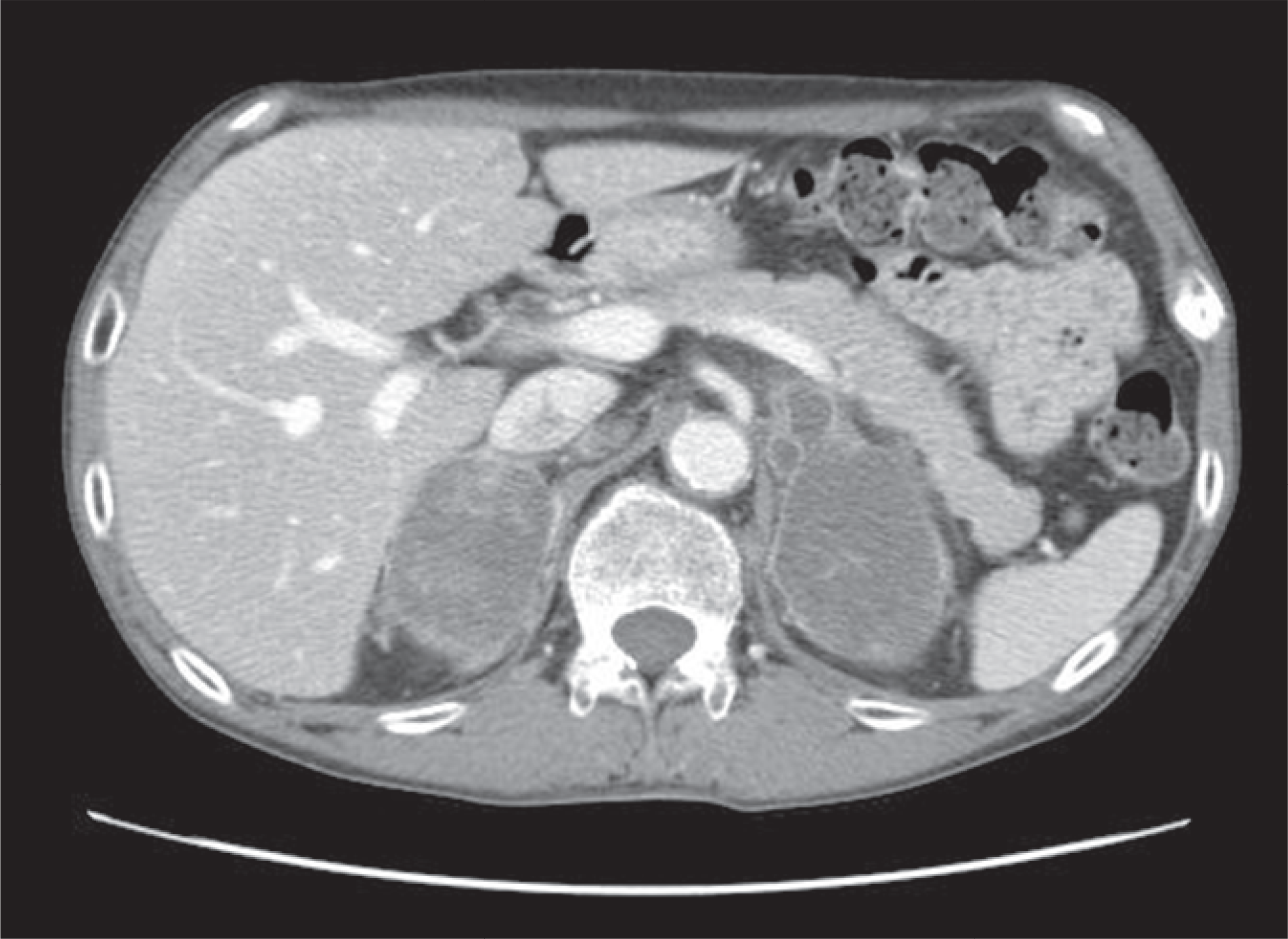
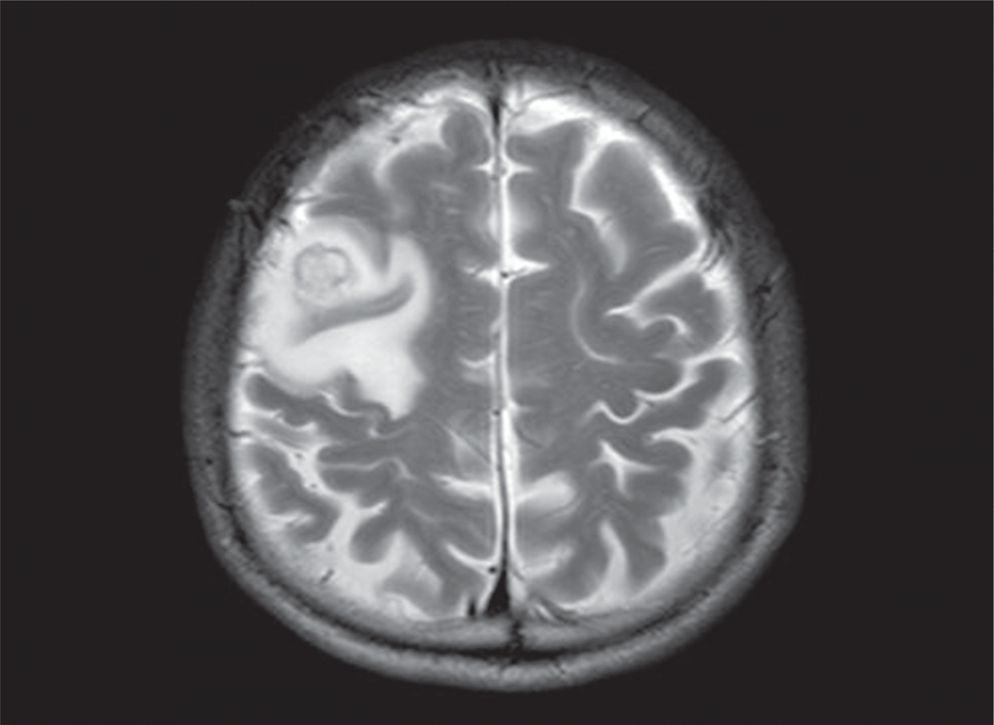
Adrenocortical carcinoma is often functional and presents with signs and symptoms of adrenal steroid hormone excess. Adrenal insufficiency secondary to bilateral adrenocortical carcinoma is a particularly rare complication. We recently encountered a case of bilateral adrenocortical carcinoma complicated by adrenal insufficiency. A 52-year-old male was transferred to this hospital com-plaining of general weakness and weight loss. A bilateral adrenal mass was detected on abdomen CT. Plasma cortisol and aldoste-rone failed to rise during the rapid ACTH stimulation test. The CT-guided adrenal biopsy revealed findings consistent with adrenocortical carcinoma. Left hemiparesis was developed and brain metastasis was detected via brain MRI. Despite the application of gamma knife surgery and chemotherapy, the disease progressed and the patient died.
REFERENCES
1. Luton JP, Cerdas S, Billaud L, Thomas G, Guilhaume B, Bertagna X, Lau-dat MH, Louvel A, Chapuis Y, Blondeau P, Bonnin A, Bricaire H. Clinical features of adrenocortical carcinoma, prognostic factors, and the effect of mitotane therapy. N Engl J Med. 322:1195–1201. 1990.

2. Kasperlik-Zaluska AA, Migdalska BM, Zgliczynski S, Makowska AM. Adrenocortical carcinoma. A clinical study and treatment results of 52 patients. Cancer. 75:2587–2591. 1995.

3. Yoon CH, Jung TS, Jung HS, Lee EY, Bae SJ, Kim JY, Chung JH, Min YK, Lee MS, Lee MK, Kim KW. Analysis of clinical features of Korean patients with adrenocortical carcinoma. J Korean Soc Endocrinol. 21:47–52. 2006.

4. Wajchenberg BL, Albergaria Pereira MA, Medonca BB, Latronico AC, Campos Carneiro P, Alves VA, Zerbini MC, Liberman B, Carlos Gomes G, Kirschner MA. Adrenocortical carcinoma: clinical and laboratory observations. Cancer. 88:711–736. 2000.
5. Koch CA, Pacak K, Chrousos GP. The molecular pathogenesis of heredi-tary and sporadic adrenocortical and adrenomedullary tumors. J Clin Endocrinol Metab. 87:5367–5384. 2002.

6. Koschker AC, Fassnacht M, Hahner S, Weismann D, Allolio B. Adrenocortical carcinoma – improving patient care by establishing new structures. Exp Clin Endocrinol Diabetes. 114:45–51. 2006.

7. Wooten MD, King DK. Adrenal cortical carcinoma. Epidemiology and treatment with mitotane and a review of the literature. Cancer. 72:3145–3155. 1993.

8. Icard P, Goudet P, Charpenay C, Andreassian B, Carnaille B, Chapuis Y, Cougard P, Henry JF, Proye C. Adrenocortical carcinomas: surgical trends and results of a 253-patient series from the French Association of Endocrine Surgeons study group. World J Surg. 25:891–897. 2001.

9. Allolio B, Hahner S, Weismann D, Fassnacht M. Management of adrenocortical carcinoma. Clin Endocrinol (Oxf). 60:273–287. 2004.

10. Cho WY, Park NC. Bilateral nonfunctioning adrenocortical carcinoma: a case report. Korean J Urol. 32:1024–1027. 1991.
11. King DR, Lack EE. Adrenal cortical carcinoma: a clinical and pathologic study of 49 cases. Cancer. 44:239–244. 1979.

12. Foster M, Nolan RL, Hong HH. Bilateral primary adrenocortical carcinoma complicated by Addisonian crisis: case report. Can Assoc Radiol J. 52:220–222. 2001.
13. Grumbach MM, Biller BM, Braunstein GD, Campbell KK, Carney JA, Godley PA, Harris EL, Lee JK, Oertel YC, Posner MC, Schlechte JA, Wie-and HS. Management of the clinically inapparent adrenal mass (“inciden-taloma”). Ann Intern Med. 138:424–429. 2003.

14. Yun M, Kim W, Alnafisi N, Lacorte L, Jang S, Alavi A. 18F-FDG PET in characterizing adrenal lesions detected on CT or MRI. J Nucl Med. 42:1795–1799. 2001.
15. Maurea S, Mainolfi C, Bazzicalupo L, Panico MR, Imparato C, Alfano B, Zivi-ello M, Salvatore M. Imaging of adrenal tumors using FDG PET: comparison of benign and malignant lesions. AJR Am J Roentgenol. 173:25–29. 1999.

16. Szolar DH, Korobkin M, Reittner P, Berghold A, Bauernhofer T, Trummer H, Schoellnast H, Preidler KW, Samonigg H. Adrenocortical carcinomas and adrenal pheochromocytomas: mass and enhancement loss evaluation at delayed contrast-enhanced CT. Radiology. 234:479–485. 2005.

17. Lam KY, Lo CY. Metastatic tumours of the adrenal glands: a 30-year experience in a teaching hospital. Clin Endocrinol (Oxf). 56:95–101. 2002.

19. Fishman EK, Deutch BM, Hartman DS, Goldman SM, Zerhouni EA, Siegelman SS. Primary adrenocortical carcinoma: CT evaluation with clinical correlation. AJR Am J Roentgenol. 148:531–535. 1987.

20. Chiche L, Dousset B, Kieffer E, Chapuis Y. Adrenocortical carcinoma ex-tending into the inferior vena cava: presentation of a 15-patient series and review of the literature. Surgery. 139:15–27. 2006.

21. Kandpal H, Sharma R, Gamangatti S, Srivastava DN, Vashisht S. Imaging the inferior vena cava: a road less traveled. Radiographics. 28:669–689. 2008.

22. Allolio B, Fassnacht M. Clinical review: Adrenocortical carcinoma: clinical update. J Clin Endocrinol Metab. 91:2027–2037. 2006.
23. Berruti A, Terzolo M, Sperone P, Pia A, Casa SD, Gross DJ, Carnaghi C, Casali P, Porpiglia F, Mantero F, Reimondo G, Angeli A, Dogliotti L. Eto-poside, doxorubicin and cisplatin plus mitotane in the treatment of advanced adrenocortical carcinoma: a large prospective phase II trial. Endocr Relat Cancer. 12:657–666. 2005.

24. Terzolo M, Angeli A, Fassnacht M, Daffara F, Tauchmanova L, Conton PA, Rossetto R, Buci L, Sperone P, Grossrubatscher E, Reimondo G, Bollito E, Papotti M, Saeger W, Hahner S, Koschker AC, Arvat E, Ambrosi B, Loli P, Lombardi G, Mannelli M, Bruzzi P, Mantero F, Allolio B, Dogliotti L, Berruti A. Adjuvant mitotane treatment for adrenocortical carcinoma. N Engl J Med. 356:2372–2380. 2007.


 PDF
PDF ePub
ePub Citation
 Print
Print


 XML Download