

 PDF
PDF ePub
ePub Citation
 Print
Print


INTRODUCTION
The Wnt family of proteins plays essential roles conserved from flies to human in many developmental processes including cell fate specification, polarity, migration and proliferation. It has becoming increasingly clear that Wnt proteins activate multiple intracellular signaling cascades to achieve the diverse biological functions. One such cascade that has been most intensely studied to date involves the multi-functional protein β-catenin; this cascade is commonly known as the canonical Wnt pathway. In this pathway, extracellular Wnt proteins (19 members in mammals) engage both the low-density lipoprotein receptor-related proteins (LRP-5 and LRP-6) [18, 22,27,31] and the Frizzled family of membrane receptors (11 members in mammals), resulting in stabilization of the cytosolic β-catenin through inhibition of GSK3β [13,32]. The stabilized β-catenin accumulates and transports into the nucleus where it interacts with transcription factors including lymphoid enhancer-binding factor-1 (Lef-1) and T cell factors (Tcf1-4), leading to transcriptional activation of downstream target genes [7].
For nearly a decade, bone biologists have studied Wnt signaling intensely. This strong interest was kindled by the discovery that loss-of-function mutations in the Wnt co-receptor LRP-5 causes the hereditary human disease osteoporosis-pseudoglioma syndrome [8]. Further supporting the role of LRP5 in bone mass regulation, multiple gain-of-function mutations in LRP5 were found to cause a high-bone-mass syndrome in humans [3,16]. Moreover, loss-of-function mutations in a secreted Wnt antagonist sclerostin (SOST) were discovered to cause rare high-bone-mass syndromes known as sclerosteosis and the van Buchem disease [1,2,4,25]. The role of Wnt signaling in bone formation, as revealed by the human genetic studies, was further demonstrated in mouse genetic models. Specifically, mice deficient in Lrp-5(Lrp-5-/-) exhibit low bone mass postnatally due to reduced osteoblast proliferation and function [14]. Moreover, mouse embryos lacking β-catenin in the skeletal progenitors failed to form osteoblasts altogether [6,11,12,23]. Overall, genetic evidence from both human and mouse strongly support the notion that Wnt signaling positively regulates bone formation.
NONCANONICAL WNT SIGNALING AND OSTEOBLAST DIFFERENTIATION
In addition to the β-catenin-mediated pathway, Wnt signaling through β-catenin-independent mechanisms, collectively termed noncanonical pathways, also plays fundamental roles in the vertebrate embryo [29]. Most notably, a β-catenin-independent pathway that involves the Rac1 and Cdc42 small GTPases, as well as the downstream kinase JNK, has been implicated in the directed migration and intercalation of cells towards the midline during gastrulation, a process known as the convergent extension (CE) movement [5,9,10,20]. This pathway is often considered to be similar to the planar cell polarity (PCP) pathway extensively studied in Drosophila. In addition, studies in Xenopus embryos have shown that Wnt proteins can activate heterotrimeric G-proteins and lead to calcium mobilization within the cell [15]. This process, often known as the "Ca++ pathway", is sensitive to pertussis toxin and therefore most likely mediated through the Gi subfamily of α subunits. Although the vast majority of studies on non-canonical Wnt signaling have been on its role on cell polarity and directed movement, recent evidence has revealed that β-catenin-independent mechanisms also play significant roles in osteoblast differentiation.
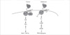
In an effort to uncover downstream effectors through which Wnt3a induces osteoblast differentiation, we serendipitously discovered a PKCδ-dependent pathway that contributes to Wnt-induced bone formation [28] (Fig. 1). By comparing the protein profiles of ST2 cells treated with Wnt3a versus untreated cells by proteomics analyses, we found that the protein MARCKS (meristoylated alanine-rich C kinase substrate), a prototypic PKC substrate, was robustly phosphorylated by Wnt3a signaling. Subsequent studies identified PKCδ as the critical PKC isoform that mediates the Wnt-induced phosphorylation. More importantly, specific knockdown of PKCδ inhibited Wnt-induced osteoblast differentiation, and PKCδ-null mouse embryos exhibited a notable delay in bone collar formation. We further investigated the upstream mechanism through which Wnt3a activates PKCδ and found that heterotrimeric G-proteins composed of the Gq subfamily of α subunits are important. Because this pathway is insensitive to pertussis toxin (which inhibits Gi but not Gq subfamily), it therefore represents a separate G-protein-mediated pathway distinct from the aforementioned Ca++ pathway. Adding to the physiological significance of this mechanism, we found that Wnt7b, a ligand selectively expressed in osteogenic cells in vivo, indeed activates the PKCδ pathway but not the β-catenin pathway, and nonetheless induces osteoblast differentiation in cell cultures. Thus, this study has provided evidence that a PKCδ-dependent mechanism, contributes to the osteogenic function of Wnt proteins independent of β-catenin. The critical target for PKCδ however is currently unknown.
More recently, Takada et al. [26] discovered that non-canonical signaling by Wnt5a promotes osteoblast differentiation while inhibiting adipogenesis from ST2 cells (Fig. 1). In particular, the authors showed that Wnt5a activates a kinase cascade CaMKII-TAK1-NLK which leads to phosphorylation of a histone methyltransferase that suppress PPARγ-target gene activation through inhibitory histone methylation of the promoters. Due to the suppression of the adipocyte fate, the authors argue that osteoblast differentiation is enhanced from the bi-potential mesenchymal progenitor. Because CaMKII (calcium/calmodulin-dependent protein kinase II) is activated by Ca++, the kinase cascade uncovered here could be a part of the Ca++ pathway as discussed above. This connection however was not directly demonstrated in this study. Overall, this study has revealed a mechanism through which Wnt proteins can alter gene transcription and hence cell fate decisions through a β-catenin-independent mechanism.
RAC1-JNK2 SIGNALING AS A CRITICAL COMPONENT OF CANONCIAL WNT SIGNALING
Although nuclear localization of β-catenin is a prerequisite for canonical Wnt signaling, the underlying mechanism is poorly understood. In a prevailing conventional view, β-catenin is stabilized in the cytoplasm to a threshold level that allows passive entry into the nucleus. A recent study from our group however challenges this view, and provides evidence that nuclear localization is a necessary regulatory step. In particular, Rac1 and JNK2 activation appears to be required for β-catenin nuclear localization and signaling [33].
Perhaps the most surprising aspect of this finding is that the Rac1-JNK signaling module, conventionally believed to function exclusively in a non-canonical Wnt pathway, performs a critical function in β-catenin signaling. While the role of Rac1-JNK in PCP and CE is well established, the new finding highlights the realization that the same signaling molecules may perform different roles in the cell, depending on their specific interaction partners and whether they are activated in a particular protein complex. Indeed, we found that β-catenin is present in a protein complex with Rac1 and JNK1/2 in the cytoplasm of ST2 cells, and Wnt3a selectively activates JNK2 in the complex. More importantly, Wnt7b, which does not activate β-catenin signaling even though it stabilizes the protein, does not activate JNK2 in the Rac1-JNK1/2-β-catenin protein complex, despite the fact that JNK2 activation can be detected in the whole cell lysate.
The role of Rac1 and JNK2 in β-catenin signaling has been confirmed by others in a colon adenoma model [21]. There, the authors found that loss of the tumor suppressor APC (adenomatous polyposis coli), which robustly stabilizes β-catenin, is insufficient for causing β-catenin nuclear localization, whereas addition activation of KRAS is effective. The authors further showed that KRAS activated Rac1 and JNK2, and inhibition of the latter two molecules abolished β-catenin nuclear localization and signaling in these cancer cells. Thus, the colon adenoma cancer cells appear to have hijacked the same mechanisms used by normal Wnt signaling.
RETHINKING CANONICAL VERSUS NONCANONICAL WNT PATHWAYS
Although "canonical" versus "noncanonical" Wnt pathways have been customarily used in the Wnt field, the new evidence calls for an understanding of these terms. First, it has become clear that the same Wnt ligand can activate multiple intracellular pathways in the same cell. This was evident in our studies where Wnt3a activates both the β-catenin and the PKCδ pathway [28]. The divergence in intracellular signaling pathways likely occurs at the level of cell-surface receptors, including both the Frizzled family members and the atypical receptor kinase Ryk [17,24], as well as the orphan receptor tyrosine kinase Ror2 [19]. Second, the same Wnt ligand can activate different pathways in different cells. For instance, we've shown that Wnt7b fails to activate β-catenin signaling in ST2 cells [28,33], but others have shown that it can activate the pathway in epithelial and vascular smooth muscle cells [30]. The difference could be caused by the presence of different receptors in different cells. Third, whereas β-catenin stabilization is clearly important, it is not synonymous with β-catenin signaling in the nucleus. Indeed, the additional regulatory steps for β-catenin signaling may well be "non-canonical", as demonstrated by the involvement of Rac1 and JNK2 [33]. Finally, whereas "canonical" is defined as β-catenin-dependent, the broad category of "non-canonical pathways" may include an unknown number of cascades that remain to be discovered. Thus, when it comes to Wnt signaling, one would be wise to keep a very open mind for the years to come.
 XML Download