

 PDF
PDF ePub
ePub Citation
 Print
Print


INTRODUCTION
Fasting plasma glucose (FPG) should be measured for the screening or diagnosis of impaired fasting glucose and diabetes (hyperglycemic states) [12]. The standard guidelines for blood sample handling indicate that plasma or serum must be separated from cells as soon as possible and within a maximum of 2 hours [3]. Therefore, the serum glucose concentrations are often measured with automatic analyzers in health examinations, and these are used as a screening test for glycemic states [4]. This may be one of the main reasons for the diagnosis of hyperglycemic states to be underestimated in Korea.
It is well known that the glucose concentrations decrease over time in whole blood ex vivo as a result of glycolysis [5]. However, no recent reports have addressed the level of the difference between the fasting serum glucose and FPG in Korea. The purpose of this study was to compare the fasting serum glucose level with the FPG for diagnosing hyperglycemic states in real-life clinical situations. We also evaluated an ordinary time delay in sample processing using our clinical laboratory tests to assess health examinations at the National Health Insurance Corp. for a period of a month to determine how time delays impact the diagnoses of hyperglycemic states.
METHODS
We recruited 1,254 participants who were diagnosed with normoglycemia or IFG using the serum blood glucose level; these were from 2,028 participants enrolled in Hallym University Sacred Heart Hospital and Kangbuk Samsung Hospital for evaluating metabolic abnormalities, including IFG or dyslipidemia according to health examinations performed by the National Health Insurance Corp. or by personal medical checkup between 2006 and 2009.
Fasting blood samples were collected from all 1,254 participants at our centers in the morning after a 12-hour overnight fast. In two consecutive samples, 3 mL of blood was drawn and placed in common tubes for the glucose measurement. Serum samples were commonly assembled at room temperature and centrifuged within 1 hour. The fasting serum glucose level was measured within 2 hours with an automated analyzer using the hexokinase/glucose-6-phosphate dehydrogenase method (Modular DPE; Hitachi Hitechnologies, Tokyo, Japan). The plasma samples were immediately centrifuged and the FPG was measured with an enzymatic assay (BIOSEN C-line; EKF Diagnostics GmbH, Berlin, Germany) within 15 minutes. We also investigated a time delay from blood sampling to glycemic testing using the order communication system (RefoMax, Seoul, Korea) at Hallym University Sacred Heart Hospital as part of health examinations performed by the National Health Insurance Corp. for a period of month because we could not directly measure the real serum-clot contact time for clinical chemistry laboratory results. We conducted a cross-sectional study, which was approved by the Institutional Review Board of Hallym University Sacred Heart Hospital.
RESULTS
A total of 2,028 participants were initially included in this study. Of these, 717 with newly diagnosed diabetes and 57 with no biochemical data were excluded; as a result, 1,254 subjects were finally included in this analysis.
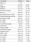
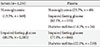
The characteristics of the study population are presented in Table 1. Their mean glucose concentrations were 119.4±9.9 mg/dL for plasma and 108.5±6.5 mg/dL for serum (mean difference, 10.9±7.4 mg/dL). Overall, 1,254 subjects were diagnosed with normoglycemia (n=169, 13.5%) or IFG (n=1,085, 86.5%) through measuring the fasting serum glucose at our centers. In this study population (n=1,254), 20.9% had newly diagnosed diabetes according to their FPG levels. Of the participants with normoglycemia (n=169), 105 (62.1%) and 24 (14.2%) were newly diagnosed with IFG and diabetes, respectively, based on their FPG levels (Table 2).
The time delay from blood sampling to glycemic testing at our clinical laboratory for health examinations of the National Health Insurance Corp. averaged 78±52 minutes for 729 subjects for the fasting serum glucose level of the 844 subjects who were examined over a period of 1 month.
DISCUSSION
The results of this study showed that the serum glucose measurement and ordinary time delay in the sample processing before getting the result of that measurement may underestimate the diagnosis of hyperglycemic states in real-life clinical situations. Decreases in the glucose concentrations in whole blood ex vivo have been reported to average 5% to 7% (10 mg/dL) per hour [5]. We performed a preliminary study of the serum-clot contact effect in 33 tests. The blood samples were kept at room temperature before serum-clot separation, and glucose concentrations were measured for 3 hours at 30-minute intervals. The serum glucose level with each processing delay of 30 minutes decreased by an average of 7.4±2.2 mg/dL per hour, and the largest decrease (4.2±1.1 mg/dL) occurred in the first 30 minutes. This decrease in the glucose concentration may result in the missed diagnosis of hyperglycemic states for a large proportion of the population with glucose levels near the cutoffs for these diagnoses [6]. The importance of early diagnosis for diabetes, which facilitates prevention of diabetic complications, has been emphasized in Korea because its rate of diabetes-related deaths is highest among the Organization for Economic Co-operation and Development (OECD) nations even though significant progress has been made in reducing the cardiovascular risk factors of Korean patients with diagnosed diabetes [7].
Several studies have demonstrated that the glucose concentrations are slightly higher in plasma than in serum [8910], while others have found no significant difference between these levels [1112]. However, these results do not seem to reflect the real clinical settings because the studies were conducted according to study protocols. We have confirmed that time delays (average of 78±52 minutes) occurred in the process of measuring the serum glucose level at our clinical laboratory for National Health Examinations, and this delay is considered to be one of the important reasons for the diagnosis of hyperglycemic states to be underestimated in Korea. A previous study from the Korea National Health and Nutrition Examination Survey also showed that serum separation and refrigeration within 30 minutes of venous sampling is recommended over the NaF (non-adjacent form) method to minimize the pre-analytical impact on diabetes detection [1314].
There are several limitations in our study. First, because all of our recruited subjects had visited hospitals, there may have been a selection bias toward higher risk factors for diabetes. Second, our cross-sectional study results may be somewhat limited due to the lack of prospective follow-up data, limiting the clinical implications of our findings. Despite these limitations, the present study, to the best of our knowledge, is the first report to show the direct comparison between the fasting serum glucose and FPG levels measured at approximately the same time when diagnosing hyperglycemic states. Glucose concentrations should be measured in plasma or serum immediately after sample separation (or within a maximum of 30 minutes) for early diagnosis and timely intervention in hyperglycemic states. Further prospective studies are also necessary to compare the fasting serum glucose level with the FPG to diagnose hyperglycemic states and evaluate the ordinary delay in sample processing in the Korean population.
 XML Download