Journal List > Diabetes Metab J > v.40(2) > 1084872

Yoon, Ha, Kim, Moon, Choi, Lim, Park, Lim, Kim, Park, and Jang: Hyperglycemia Is Associated with Impaired Muscle Quality in Older Men with Diabetes: The Korean Longitudinal Study on Health and Aging

Abstract

Background

The study aimed to investigate the influence of hyperglycemia on muscle quality in older men with type 2 diabetes.

Methods

This was a subsidiary study of the Korean Longitudinal Study of Health and Aging. Among 326 older men consenting to tests of body composition and muscle strength, 269 men were ultimately analyzed after the exclusion because of stroke (n=30) and uncertainty about the diagnosis of diabetes (n=27). Body composition was measured using dual-energy X-ray absorptiometry and computed tomography. Muscle strength for knee extension was measured using an isokinetic dynamometer. Muscle quality was assessed from the ratio of leg strength to the entire corresponding leg muscle mass.

Results

The muscle mass, strength, and quality in patients with type 2 diabetes did not differ significantly from controls. However, when patients with diabetes were subdivided according to their glycemic control status, patients with a glycosylated hemoglobin (HbA1c) level of ≥8.5% showed significantly decreased leg muscle quality by multivariate analysis (odds ratio, 4.510; P=0.045) after adjustment for age, body mass index, smoking amount, alcohol consumption, physical activity, and duration of diabetes. Physical performance status was also impaired in subjects with an HbA1c of ≥8.5%.

Conclusion

Poor glycemic control in these older patients with diabetes was associated with significant risk of decreased muscle quality and performance status. Glycemic control with an HbA1c of <8.5% might be needed to reduce the risk of adverse skeletal and functional outcomes in this population.

INTRODUCTION

A progressive decline in muscle mass and strength, termed sarcopenia, develops as a consequence of aging [1]. The prevalence of sarcopenia differs depending on the definition and methods of assessment; it ranges from 8% to 40% of adults aged over 60 years [2]. Sarcopenia results in frailty, loss of independence, physical disability, and increased mortality in older adults [34]. Diabetes also has been associated with an increased risk of developing physical disability in older adults [56]. Chronic conditions, such as visual disturbance, diabetic complications, comorbidities, and depression, have been known to be associated with physical disability in patients with diabetes; however, these accounted for only some of the impairments [78]. Given the relationship between decreased muscle strength or quality and physical disability in older adults [49], it is very important to study whether decreases in muscle strength and quality occur in older people with diabetes. Although lower extremity weakness is a common complaint in patients with diabetes and poor glycemic control seen in clinical practice, few studies have investigated the effects of glycemic control on muscle strength and quality.
In this study, we investigated whether poor glycemic control had an impact on muscle strength and quality in older men with diabetes among a community-based cohort enrolled in the Korean Longitudinal Study of Health and Aging (KLoSHA). Possible relevant factors that could affect muscle performance, such as adipocytokines, insulin resistance, and perimuscular fat amounts, were also evaluated.

METHODS

Study design and subjects

KLoSHA is a community-based cohort covering 1,000 patients (439 men and 561 women), aged 65 years or older, first recruited in 2005. The population and study details for this cohort have been published previously [10]. Among these patients, 326 men agreed to have their body composition and muscle strength tested. Patients with a history of cerebrovascular accidents (n=30) were excluded because they might have muscle weakness and/or loss of muscle mass caused by denervation or inactivity. In addition, 27 patients with diabetes mellitus who had glycosylated hemoglobin (HbA1c) levels of <6% were excluded. Ultimately, 269 men were evaluated.
A normal control group was defined as comprising patients who did not meet the diagnostic criteria of diabetes mellitus following a 75-g oral glucose tolerance test and an HbA1c value of <6.5%. Diabetes mellitus was defined using diagnostic criteria recommended by the American Diabetes Association [11] or by the current use of insulin or oral hypoglycemic medication. Patients with diabetes were subdivided into four groups according to their HbA1c values. The Institutional Review Board of Seoul National University Bundang Hospital approved this study (IRB B-0706/046-012). Written informed consent was obtained from every patient.

Medical histories and anthropometry

Each patient's medical history, including diseases and medications and personal details, such as alcohol intake, smoking habit, and physical activity level, was investigated by trained nurses, who were certified in epidemiology and the assessment of elderly patients. A physical activity score was assessed as described previously [12].
Height and body weight were measured in patients wearing light clothing while barefoot and used to calculate the body mass index (BMI). Waist circumference was measured at the narrowest point between the lower limit of the ribcage and the iliac crest. Hip circumference was measured as the maximal circumference over the buttocks.

Body composition measurement

Body composition was measured by dual-energy X-ray absorptiometry (DXA; Lunar Corp., Madison, WI, USA). Appendicular skeletal muscle mass (ASM) was calculated as the sum of the lean soft tissue mass in the arms and legs. The ab-dominal adipose tissue areas were quantified by computed tomography (CT) scan at a 90-kV exposure (Somatom Sensation 16; Siemens, Munich, Germany). A 10-mm CT slice scan was acquired at the umbilical level to measure abdominal and visceral fat areas by measuring the mean value of all pixels within the range of –190 to –50 Hounsfield units. A CT scan at the mid-thigh level between the pubic symphysis and inferior condyle of the femur was performed. Measurements of the cross-sectional mid-thigh fat areas (subcutaneous and intermuscular fat areas) were obtained by measuring the mean value of all pixels within the range of –190 to –50 Hounsfield units.

Muscle strength and quality measurements

Muscle strength was measured using an isokinetic dynamometer (Biodex System 3 Pro; Biodex Inc., Shirley, NY, USA) for knee extension. The maximal voluntary isokinetic torque was assessed in Newton meters at an angular velocity of 60°/sec. At least three, but no more than six, maximal efforts were allowed to produce three overlying curves, and the mean maximal torque was determined and used for the analysis. The dominant leg was used unless the subject experienced pain in it. Muscle quality was expressed as the ratio of the strength measured to the entire corresponding leg muscle mass in kilograms as measured by DXA.

Assessment of physical performance status

Lower extremity physical performance was assessed using the Short Physical Performance Battery (SPPB) method. This consists of three subtests for balance, walking, and muscle strength. The SPPB summary performance score (range, 0 to 12) is the sum of individual test scores, with higher scores indicating better lower extremity performance [13]. The SPPB approach has been reported as a strong, independent predictor of physical disability, institutionalization, and mortality in older adults [14].

Biochemical parameters

In all patients, plasma glucose and insulin concentrations were measured after a 12-hour fast. A 75-g oral glucose tolerance test was administered to patients who had not been diagnosed with diabetes. Plasma glucose concentration was measured using the glucose oxidase method. Plasma insulin concentration was measured by radioimmunoassay (Linco Research, St. Charles, MO, USA). The homeostasis model assessment of insulin resistance (HOMA-IR) was calculated [15]. HbA1c concentrations were measured using ion-exchange high-performance liquid chromatography (VARIANT II; Bio-Rad Laboratories, Hercules, CA, USA). Total cholesterol, triglycerides, high density lipoprotein, and low density lipoprotein cholesterol levels were measured enzymatically using an autoanalyzer (Hitachi 747; Hitachi Ltd., Tokyo, Japan). To evaluate inflammatory status, high-sensitivity C-reactive protein (hs-CRP) concentrations were measured using immunonephelometry (Dade Behring, Marburg, Germany). Plasma adiponectin and retinol binding protein 4 (RBP4) levels were measured using enzyme-linked immunosorbent assay kits (AdipoGen, Seoul, Korea).

Statistical analyses

All data are presented as the mean and standard deviation and were analyzed using SPSS version 20.0 (IBM Co., Armonk, NY, USA). The baseline characteristics, muscle mass, strength, and quality of test and normal control groups were compared using Student t-tests or chi-square tests. HOMA-IR values were skewed, so they were normalized by logarithmic transformation for analyses. Comparisons of muscle mass, strength, quality, and performance status between subgroups were assessed with one-way analysis of variance. Tukey's post hoc test was performed when a significant difference was observed with the analysis of variance. Multiple logistic regression analysis was used to determine the independent effect of glycaemic status on muscle quality. P<0.05 was considered statistically significant.

RESULTS

Characteristics of patients according to their glycemic control status

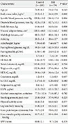
Among the 269 men, 79 (29.4%) had type 2 diabetes mellitus (T2DM), of whom 33 were newly diagnosed. Subjects with T2DM had a higher BMI and systolic blood pressure (Table 1). In terms of body composition, total body fat mass and visceral adipose tissue areas were significantly greater in patients with T2DM, whereas mid-thigh fat area and ASM did not differ between two groups. The ASM/height2 and frequency of sarcopenia (ASM/height2 <6.43 kg/m2) were also not different between two groups. The mean fasting glucose, HbA1c, and HOMA-IR were significantly higher in subjects with T2DM compared with control subjects. Lipid profiles, serum creatinine, and hs-CRP levels were not different between two groups. In addition, lifestyle parameters of alcohol consumption, smoking amount, and physical activity score were not different. Serum adiponectin concentration was significantly lower in patients with T2DM, whereas serum RBP4 concentration was not.

Comparisons of lower extremity muscle mass, strength, and quality, according to the glycemic status

There was no difference of muscle mass, strength, and quality of low extremity between subjects with T2DM and normal controls (Table 1). We subdivided patients with T2DM into four groups according to HbA1c level to evaluate whether there was any difference in lower extremity muscle mass and strength according to the level of glycemic control. The duration of T2DM in patients with HbA1c ≥6.5% was longer than those with HbA1c <6.5%, but no significant differences of muscle-related indices, including leg lean body mass and knee peak torque extension, were observed among the groups. However, lower extremity muscle quality was significantly different between groups; in post hoc analysis, only the group with HbA1c ≥8.5% showed significantly decreased muscle quality compared with normal controls and patients with diabetes and an HbA1c of <8.5% (Fig. 1).

Multivariate analysis for the risk of decreased muscle quality related to glycemic control

Multivariate analysis was applied to investigate whether poor glycemic control had an independent effect on decreased muscle quality. In a correlation analysis, HOMA-IR, adiponectin, RBP4, and mid-thigh fat area did not show any significant relationship with muscle quality and were excluded from the multivariate analysis. When the muscle quality belonged to the lowest quartile of the study population, it was defined as decreased muscle quality. After adjustment for age, BMI, smoking amount, alcohol consumption, physical activity score, and duration of diabetes, older patients with diabetes and HbA1c level of ≥8.5% had a higher risk of having decreased muscle quality; the odds ratio was 4.540 when compared with normal control patients (Table 2).

Physical performance status related to glycemic control

The SPPB score was significantly lower in patients with HbA1c of ≥8.5% than in patients with HbA1c of <8.5% and in normal controls, which means that a decrease in muscle quality related to poor glycemic control was also related to functional impairment in older adults (Fig. 2).

DISCUSSION

In this study, merely the presence of T2DM did not cause declines in muscle mass, strength, or quality in older men. However, patients with diabetes and poor glycemic control (defined as HbA1c level of ≥8.5%) showed significantly decreased lower extremity muscle quality. Statistical significance was maintained in the analysis even after adjusting for age, BMI, smoking habit, alcohol use, and exercise status. Poor glycemic control status was also associated with functional impairments as estimated by SPPB scores. These results are consistent with those of Park et al. [16], which showed decreased muscle quality in older patients with diabetes. However, in our study, the presence of diabetes itself was not associated with a decline in muscle quality. Most patients with diabetes in this study had good glycemic control (the mean HbA1c level was 7.2%), and this might have obscured the influence of glycemic status on muscle. Oxidative stress and the accumulation of advanced glycation end products have been suggested as mechanisms leading to sarcopenia [11718]. There are several other mechanisms that could explain why poor glycemic control might lead to a decline in muscle quality, such as decreased glucose utilization by muscle [19], increased levels of systemic inflammatory cytokines such as interleukin-6, tumor necrosis factor alpha, and hs-CRP [2021], neuropathic processes involving motor neurons [22], and mitochondrial dysfunction [23]. In this study, we evaluated adipocytokines, insulin resistance, and perimuscular fat as possible additional factors related to decreased muscle quality. However, we found no significant relationship between these variables and muscle quality.
Muscle quality, defined as muscle strength per unit muscle mass, has been used to assess muscle function and was considered a more meaningful indicator of muscle function than strength alone in previous studies [2425]. It has been demonstrated that aging itself can lead to deteriorating muscle quality or to a disproportionate decrease in muscle strength compared with a decrease in muscle mass [26]. In our study, men with diabetes and poor glycemic control showed significantly lower muscle quality and poor performance scores. Our study results indicate that poor glycemic control in such older men could exacerbate age-related muscle quality deterioration.
Sarcopenia is related to difficulties in rising from a chair [27], slow gait, balancing problems, and falls [28], which are important components of functional performance. To test whether decreased muscle quality associated with poor glycemic control might influence physical performance, SPPB scores were compared between groups: patients with diabetes with HbA1c levels of 8.5% or higher who showed decreased muscle quality also had a significantly lower SPPB score. Our results also supported the glycemic treatment goal suggested by the American Diabetes Association [29].
There were several limitations to this study. First, the cross-sectional design limited any interpretation of the causal relationship between glycemic control and muscle quality. Thus, it is unclear whether poor glycemic control decreases muscle quality or poor quality muscle affects glycemic control. This needs to be verified in future studies using a prospective design. Second, instead of intermuscular fat area in the mid-thigh, we measured a combined area that included both subcutaneous and intermuscular fat. The lack of correlation between perimuscular fat area and muscle quality could have been associated with the confounding effect of the subcutaneous fat area. Third, KLoSHA was a prospective cohort comprising 1,000 randomly sampled, community-dwelling, older adults. Therefore, there was uneven distribution between control and diabetes groups because the sample size determination was not performed for the comparison of muscle mass or muscle strength. Especially with the small sample size for patients with poor glycemic control, it was difficult to determine statistical significance, although there was an apparent trend according to the level of glycemic control.
Currently, there are few guidelines for target levels of glycemic control in older adults. Life expectancy, the presence of complications and comorbidities associated with diabetes, and the patient's ability and willingness to comply with a diabetes treatment program should be taken into consideration in developing a long-term management plan in older patients with diabetes. More conservative therapeutic targets have been advocated for older patients with diabetes with associated medical problems or advanced complications [30]. Given the finding in our study that poor glycemic control was associated with impaired muscle quality and functional ability in older people with diabetes, a target HbA1c level of <8.5% seems appropriate for preventing declines in muscle quality and functional performance.

Figures and Tables

Fig. 1

Poor glycemic control was associated with lower muscle quality. Older patients with diabetes and glycosylated hemoglobin (HbA1c) levels of ≥8.5% showed significantly decreased muscle quality compared with patients with HbA1c levels of <8.5%, as well as non-diabetes mellitus (DM). Nm, newton meter. aP<0.05, bP=0.053.

dmj-40-140-g001
Fig. 2

Comparison of physical performance status related to the glycemic control of older diabetic patients with glycosylated hemoglobin (HbA1c) levels of ≥8.5% showed significantly lower Short Physical Performance Battery (SPPB) scores than diabetic patients with HbA1c levels of <8.5%, as well as non-diabetes mellitus (DM). aP<0.01 vs. non-DM, bP<0.05 vs. HbA1c <8.5%.

dmj-40-140-g002
Table 1

Clinical characteristics of older men with or without type 2 diabetes mellitus

dmj-40-140-i001
Characteristic Control (n=190) DM (n=79) P valuea
Age, yr 74.9±8.5 73.4±7.4 0.143
Body mass index, kg/m2 23.5±3.2 24.8±2.9 0.002
Systolic blood pressure, mm Hg 129.9±16.2 136.4±17.8 0.004
Diastolic blood pressure, mm Hg 82.5±10.8 82.7±10.1 0.815
Body fat mass, kg 14.6±6.0 16.8±5.6 0.008
Visceral adipose tissue, cm2 120.5±64.0 152.5±62.3 0.002
Mid-thigh fat area, cm2 80.5±32.7 80.8±34.9 0.951
ASM, kg 20.2±2.8 20.6±2.7 0.139
ASM/height2, kg/m2 7.39±0.85 7.46±0.77 0.563
Fasting blood glucose, mg/dL 99.2±14.9 142.5±35.9 <0.001
Fasting insulin, µU/mL 1.70±1.66 2.43±2.10 0.217
HbA1c, % 5.7±0.3 7.2±1.0 <0.001
HOMA-IR 1.16±0.77 1.88±1.04 <0.001
Total cholesterol, mg/dL 193.0±33.3 194.5±40.5 0.758
Triglyceride, mg/dL 132.9±98.9 137.0±99.6 0.758
HDL-C, mg/dL 59.4±14.9 56.6±12.6 0.152
Creatinine, mg/dL 1.2±0.4 1.2±0.3 0.657
hs-CRP, mg/dL 0.17±0.33 0.20±0.47 0.580
Adiponectin, µg/mL 8.6±6.2 6.9±4.8 0.027
RBP4, µg/mL 61.5±29.3 63.5±28.7 0.613
Alcohol consumption, units/mo 77.5±112.6 71.8±87.3 0.782
Smoking amount, pack-year 23.8±26.6 37.9±95.8 0.223
Physical activity score 16.4±5.8 16.8±6.3 0.577
Leg lean body mass, kg 14.8±2.0 15.2±2.1 0.165
Knee peak torque extension, Nm 74.7±2.0 78.8±3.2 0.831
Knee muscle quality extension, Nm/kg 10.7±0.2 10.4±0.4 0.465
SPPB score 10.0±2.1 9.7±2.4 0.378

Values are presented as mean±standard deviation.

DM, diabetes mellitus; ASM, appendicular skeletal muscle mass; HbA1c, glycosylated hemoglobin; HOMA-IR, homeostasis model assessment of insulin resistance; HDL-C, high density lipoprotein cholesterol; hs-CRP, high-sensitivity C-reactive protein; RBP4, retinol binding protein-4; Nm, newton meter; SPPB, Short Physical Performance Battery.

aP values are for Student t-tests.

Table 2

Multivariate analysis for the risk of decreased muscle quality (<25 percentile) related to glycemic control

dmj-40-140-i002
HbA1c, % B Odds ratio 95% CI P value
<6.5 0.127 1.135 0.314–4.100 0.846
6.5–7.4 0.608 1.837 0.577–5.845 0.303
7.5–8.4 0.103 1.108 0.171–7.181 0.914
≥8.5 1.513 4.540 1.031–19.985 0.045

Adjusted for age, body mass index, smoking habit, alcohol consumption, physical activity, and duration of diabetes mellitus.

HbA1c, glycosylated hemoglobin; CI, confidence interval.

ACKNOWLEDGMENTS

This study was supported by grant from National Research Foundation grant, Ministry of Education, Science, and Technology (No. 2006–2005410), Republic of Korea.

Notes

CONFLICTS OF INTEREST No potential conflict of interest relevant to this article was reported.

References

1. Cruz-Jentoft AJ, Baeyens JP, Bauer JM, Boirie Y, Cederholm T, Landi F, Martin FC, Michel JP, Rolland Y, Schneider SM, Topinkova E, Vandewoude M, Zamboni M. European Working Group on Sarcopenia in Older People. Sarcopenia: European consensus on definition and diagnosis: report of the European Working Group on Sarcopenia in Older People. Age Ageing. 2010; 39:412–423.
2. Abellan van Kan G. Epidemiology and consequences of sarcopenia. J Nutr Health Aging. 2009; 13:708–712.
3. Fried LP, Guralnik JM. Disability in older adults: evidence regarding significance, etiology, and risk. J Am Geriatr Soc. 1997; 45:92–100.
4. Kim JH, Lim S, Choi SH, Kim KM, Yoon JW, Kim KW, Lim JY, Park KS, Jang HC. Sarcopenia: an independent predictor of mortality in community-dwelling older Korean men. J Gerontol A Biol Sci Med Sci. 2014; 69:1244–1252.
5. De Rekeneire N, Resnick HE, Schwartz AV, Shorr RI, Kuller LH, Simonsick EM, Vellas B, Harris TB. Health, Aging, and Body Composition study. Diabetes is associated with subclinical functional limitation in nondisabled older individuals: the health, aging, and body composition study. Diabetes Care. 2003; 26:3257–3263.
6. Kalyani RR, Saudek CD, Brancati FL, Selvin E. Association of diabetes, comorbidities, and A1C with functional disability in older adults: results from the National Health and Nutrition Examination Survey (NHANES), 1999-2006. Diabetes Care. 2010; 33:1055–1060.
7. Ahroni JH, Boyko EJ, Davignon DR, Pecoraro RE. The health and functional status of veterans with diabetes. Diabetes Care. 1994; 17:318–321.
8. Caruso LB, Silliman RA, Demissie S, Greenfield S, Wagner EH. What can we do to improve physical function in older persons with type 2 diabetes? J Gerontol A Biol Sci Med Sci. 2000; 55:M372–M377.
9. Kim JH, Choi SH, Lim S, Yoon JW, Kang SM, Kim KW, Lim JY, Cho NH, Jang HC. Sarcopenia and obesity: gender-different relationship with functional limitation in older persons. J Korean Med Sci. 2013; 28:1041–1047.
10. Lim S, Yoon JW, Choi SH, Park YJ, Lee JJ, Park JH, Lee SB, Kim KW, Lim JY, Kim YB, Park KS, Lee HK, Cho SI, Jang HC. Combined impact of adiponectin and retinol-binding protein 4 on metabolic syndrome in elderly people: the Korean Longitudinal Study on Health and Aging. Obesity (Silver Spring). 2010; 18:826–832.
11. American Diabetes Association. Diagnosis and classification of diabetes mellitus. Diabetes Care. 2010; Suppl 1. S62–S69.
12. Washburn RA, McAuley E, Katula J, Mihalko SL, Boileau RA. The physical activity scale for the elderly (PASE): evidence for validity. J Clin Epidemiol. 1999; 52:643–651.
13. Guralnik JM, Simonsick EM, Ferrucci L, Glynn RJ, Berkman LF, Blazer DG, Scherr PA, Wallace RB. A short physical performance battery assessing lower extremity function: association with self-reported disability and prediction of mortality and nursing home admission. J Gerontol. 1994; 49:M85–M94.
14. Guralnik JM, Ferrucci L, Simonsick EM, Salive ME, Wallace RB. Lower-extremity function in persons over the age of 70 years as a predictor of subsequent disability. N Engl J Med. 1995; 332:556–561.
15. Matthews DR, Hosker JP, Rudenski AS, Naylor BA, Treacher DF, Turner RC. Homeostasis model assessment: insulin resistance and beta-cell function from fasting plasma glucose and insulin concentrations in man. Diabetologia. 1985; 28:412–419.
16. Park SW, Goodpaster BH, Strotmeyer ES, de Rekeneire N, Harris TB, Schwartz AV, Tylavsky FA, Newman AB. Decreased muscle strength and quality in older adults with type 2 diabetes: the health, aging, and body composition study. Diabetes. 2006; 55:1813–1818.
17. Doherty TJ. Invited review: aging and sarcopenia. J Appl Physiol (1985). 2003; 95:1717–1727.
18. Jones TE, Stephenson KW, King JG, Knight KR, Marshall TL, Scott WB. Sarcopenia: mechanisms and treatments. J Geriatr Phys Ther. 2009; 32:83–89.
19. Scheck SH, Barnard RJ, Lawani LO, Youngren JF, Martin DA, Singh R. Effects of NIDDM on the glucose transport system in human skeletal muscle. Diabetes Res. 1991; 16:111–119.
20. Taaffe DR, Harris TB, Ferrucci L, Rowe J, Seeman TE. Cross-sectional and prospective relationships of interleukin-6 and C-reactive protein with physical performance in elderly persons: MacArthur studies of successful aging. J Gerontol A Biol Sci Med Sci. 2000; 55:M709–M715.
21. Visser M, Pahor M, Taaffe DR, Goodpaster BH, Simonsick EM, Newman AB, Nevitt M, Harris TB. Relationship of interleukin-6 and tumor necrosis factor-alpha with muscle mass and muscle strength in elderly men and women: the Health ABC Study. J Gerontol A Biol Sci Med Sci. 2002; 57:M326–M332.
22. Andersen H, Gadeberg PC, Brock B, Jakobsen J. Muscular atrophy in diabetic neuropathy: a stereological magnetic resonance imaging study. Diabetologia. 1997; 40:1062–1069.
23. Barazzoni R. Skeletal muscle mitochondrial protein metabolism and function in ageing and type 2 diabetes. Curr Opin Clin Nutr Metab Care. 2004; 7:97–102.
24. Metter EJ, Lynch N, Conwit R, Lindle R, Tobin J, Hurley B. Muscle quality and age: cross-sectional and longitudinal comparisons. J Gerontol A Biol Sci Med Sci. 1999; 54:B207–B218.
25. Newman AB, Haggerty CL, Goodpaster B, Harris T, Kritchevsky S, Nevitt M, Miles TP, Visser M. Health Aging And Body Composition Research Group. Strength and muscle quality in a well-functioning cohort of older adults: the Health, Aging and Body Composition Study. J Am Geriatr Soc. 2003; 51:323–330.
26. Goodpaster BH, Park SW, Harris TB, Kritchevsky SB, Nevitt M, Schwartz AV, Simonsick EM, Tylavsky FA, Visser M, Newman AB. The loss of skeletal muscle strength, mass, and quality in older adults: the health, aging and body composition study. J Gerontol A Biol Sci Med Sci. 2006; 61:1059–1064.
27. Alexander NB, Schultz AB, Warwick DN. Rising from a chair: effects of age and functional ability on performance biomechanics. J Gerontol. 1991; 46:M91–M98.
28. Wolfson L, Judge J, Whipple R, King M. Strength is a major factor in balance, gait, and the occurrence of falls. J Gerontol A Biol Sci Med Sci. 1995; 50 Spec No:64–67.
29. Kirkman MS, Briscoe VJ, Clark N, Florez H, Haas LB, Halter JB, Huang ES, Korytkowski MT, Munshi MN, Odegard PS, Pratley RE, Swift CS. Diabetes in older adults. Diabetes Care. 2012; 35:2650–2664.
30. Lebovitz HE. American Diabetes Association. Charter 31, Diabetes in older adults. Therapy for diabetes mellitus and related disorders. 5th ed. Alexandria: American Diabetes Association;2009. p. 364–365.
TOOLS
ORCID iDs

Hak Chul Jang
https://orcid.org/http://orcid.org/0000-0002-4188-6536

Similar articles