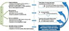
Journal List > Diabetes Metab J > v.39(4) > 1084820

This article has been corrected. See "Erratum: Figure Correction: Probiotics as Complementary Treatment for Metabolic Disorders" in Volume 39 on page 448.
Le Barz, Anhê, Varin, Desjardins, Levy, Roy, Urdaci, and Marette: Probiotics as Complementary Treatment for Metabolic Disorders

Abstract

Over the past decade, growing evidence has established the gut microbiota as one of the most important determinants of metabolic disorders such as obesity and type 2 diabetes. Indeed, obesogenic diet can drastically alter bacterial populations (i.e., dysbiosis) leading to activation of pro-inflammatory mechanisms and metabolic endotoxemia, therefore promoting insulin resistance and cardiometabolic disorders. To counteract these deleterious effects, probiotic strains have been developed with the aim of reshaping the microbiome to improve gut health. In this review, we focus on benefits of widely used probiotics describing their potential mechanisms of action, especially their ability to decrease metabolic endotoxemia by restoring the disrupted intestinal mucosal barrier. We also discuss the perspective of using new bacterial strains such as butyrate-producing bacteria and the mucolytic Akkermansia muciniphila, as well as the use of prebiotics to enhance the functionality of probiotics. Finally, this review introduces the notion of genetically engineered bacterial strains specifically developed to deliver anti-inflammatory molecules to the gut.

INTRODUCTION

Trillions of microorganisms among bacteria, archaea, viruses, and fungi inhabit our gastrointestinal tract (GIT). Bacterial cells largely outnumber our own cells, and the gut microbiota is a prolific example of a symbiotic relationship as it plays a crucial role in host's physiology and health. Studies based on gnotobiotic models and fecal microbial transplants (FMT) have provided unequivocal evidence that perturbations in bacterial communities play a key role in the pathophysiology of obesity and insulin resistance [12]. The gut microbiota is the product of a complex interaction between host's genetics and environment, and diet is one of the main driving forces shaping intestinal bacterial communities [3]. The so-called western obesogenic diet (i.e., rich in saturated/trans fat and simple sugars and poor in fibers) is associated with specific modulation of taxonomic profiles that are functionally linked with a more proinflammatory milieu and disrupted intestinal barrier. Disturbance of intestinal homeostasis then leads to excessive bacterial fragments/products internal diffusion, which promotes inflammation in key insulin-responsive tissues, resulting in insulin resistance [4].
The current knowledge suggests that gut bacterial profiles may represent new disease predictors and that manipulation of the gut microbiota could be a promising approach for the prevention and management of metabolic diseases [5]. Indeed, Cani et al. [6] were the first group to demonstrate a positive correlation between alteration of gut microbiota population, the increase of intestinal permeability and the development of metabolic endotoxemia that is characterized by the translocation of bacterial lipopolysaccharides (LPSs) into the systemic circulation and induction of inflammatory pathways in mice fed obesogenic diet.
Since Metchnikoff's era, the field of probiotics-live microorganisms that, when administered in adequate amounts, confers a health benefit on the host (Food and Agriculture Organization of the United Nations, 2002; updated by Hill et al. [7])-continues to grow thanks to the recent access to investigate the role of an increasing number of potential probiotic strains in host's physiology. According to this definition, the safety and efficacy of a given strain must be scientifically demonstrated in order to be considered as a probiotic. Here, we propose a critical review of the most recent studies concerning the effects of probiotic bacterial strains in the prevention or treatment of metabolic disorders such as obesity, insulin resistance, diabetes mellitus and its comorbidities.

MICROORGANISMS WIDELY USED AS PROBIOTICS

Most currently used probiotics belong to bifidobacteria, lactic acid bacteria (LAB), dairy propionibacteria, yeasts (Saccharomyces boulardii), Bacillus, and the gram-negative Escherichia coli strain Nissle 1917 [8]. LAB represent a heterogeneous group of microorganisms broadly present in the diet, particularly by the use of non-human strains in the fermentation of dairy products being also normal inhabitants of the gastrointestinal and urogenital tract [9]. Most of them are members of the phylum Firmicutes, while Bifidobacterium, also considered as lactic-producing bacteria, belong to Actinobacteria phylum.
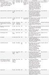
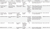
Probiotic administration has been shown to stimulate the immune response, improve lactose tolerance, help prevent diarrhea, have an anti-inflammatory effect and even restore obesity-linked gut dysbiosis [10]. Given the relationship between obesity-related disorders and gut homeostasis, probiotics may be of interest to supplement the limited arsenal of therapies against the metabolic syndrome. The diversity of reported studies in Tables 1 and 2, shows that the positive effects of probiotics are strain-specific and the idea of a "universal strain," that would provide at once all the benefits associated with probiotics, is unrealistic, even for strains of the same species [7]. In the context of obesity and metabolic disorders, probiotic supplementation may help to reduce hyperphagia [11], improving control of weight gain, fat mass loss and glucose tolerance. On the contrary, such positive effects could also be obtained without modulation of caloric intake, as demonstrated by most of the reported studies [12131415161718]. To demonstrate the beneficial effect of probiotics in improvement of metabolic disorders, researchers have access to a variety of assays such as plasma and liver cholesterol, free-fatty acids, alanine and aspartate transaminases (hepatotoxicity markers), gene and protein expressions (involved in inflammatory and metabolic pathways), etc. (Tables 1 and 2 for details).
For example, dairy products supplemented with Propionibacterium, a well-known promising non-LAB genus, may exert a probiotic effect in the colon by producing metabolites such as short-chain fatty acids (SCFA), vitamins (B8, B9, and B12), and 1,4-dihydroxy-2-naphtoic acid, bifidogenic and anti-inflammatory product (DHNA) [19]. Oksaharju et al. [20] demonstrated that Propionibacterium freudenreichii ssp. shermani JS has anti-inflammatory effects on high fat diet-induced inflammation in ApoE*3Leiden mice, with a decrease of intestinal mast cell numbers and a demonstrated intestinal but also systemic anti-inflammatory potential.
Finally, although it needs further investigation, multiple strain probiotics could confer a more effective strategy than single-strain probiotics against diet-induced obesity (DIO) [12]. Interestingly, VSL#3, a mixture of eight different strains of bacteria, has shown efficacy in prevention but also in the treatment of obesity and type 2 diabetes [11].

POTENTIAL MECHANISMS OF ACTIONS

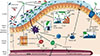
Probiotic administration is frequently associated with important shifts in gut bacterial composition, along with beneficial effects on metabolism and inflammatory tone [1617182122]. Indeed, within the gut, probiotic are in competition for nutrients, metabolites and also for antimicrobial proteins, altering gut microbiota population diversity in several ways [12]. However, it remains unclear if the modulation of gut microbiota is the cause or the consequence of probiotic treatment, or whether the mechanisms are partially or totally interdependent. The probiotic components associated with positive effects are a variety of cell constituents as polysaccharides, peptidoglycan, DNA, teichoic acids and certain cell-surface bound and secreted proteins as well as organic acids, bacteriocins, polyphosphate, and fatty acids (FA), which can modulate host responses, inhibit pathogens or interact with the intestinal microbiota [23]. Furthermore, whereas a disrupted intestinal barrier contributes to the pathogenesis of metabolic diseases, the underlying causes remain unclear. Indeed, it may include changes in nutritional factors, infections (i.e., Helicobacter pylori infection leading to an increased rate of incident diabetes), exposure to toxins, lack of exposure to microbes in early childhood, as well as impaired function and diversity of the gut microbiota [24]. Moreover, probiotic strains can not only affect the intestinal microbiota directly but also affect other organs by modulating intestinal inflammation and permeability [25]. Several potential mechanisms underlying the beneficial effects of probiotics are illustrated in Figs 1 and 2.

REDUCTION OF BODY OR LIVER FAT MASS

The "obese microbiome" is thought to display an increased capacity to harvest energy from the diet along with a decreased ability to stimulate the production of gut factors that inhibit fat deposition [1]. Furthermore, the beneficial effect of probiotics to decrease DIO is both highly strain- and model-specific [1318262728]. For example, the gut microbiota could promote storage of triglyceride in adipocytes through suppression of intestinal expression of a circulating lipoprotein lipase (LPL) inhibitor, the angiopoietin-like 4 [2]. Nevertheless, storage of excess FA is the result of unbalanced lipid absorption involving LPL and lipid catabolism [29]. In fact, Yoo et al. [12], treated DIO-mice with a combination of probiotics that resulted in a decreased expression of genes involved in FA transport and β-oxidation (Table 1). Another potential mechanism by which probiotics can counteract the negative effect of obesogenic diet is by interaction with commensal bacteria and altering expressions of microbial enzymes, especially those involved in carbohydrate metabolism or butyrate synthesis pathways [3031]. Butyrate, with acetate and propionate, are the most abundant SCFA produced by some colonic bacteria as end-products from the breakdown of non-digestible carbohydrates that pass unaffected through the small intestine [5]. Among major bacterial phyla, Bacteroidetes are recognized as acetate and propionate producers, whereas Firmicutes are more butyrate-producing bacteria. The butyrate-producer strain miyairi 588 has shown promising effects on liver homeostasis and insulin resistance in a rat model of choline-deficient diet-induced non-alcoholic fatty liver disease (NAFLD) [32]. As reported in Table 1, Ritze et al. [21] have also shown that Lactobacillus rhamnosus GG protects against NAFLD through specifically reducing liver fat mass loss in association with modulation of the carbohydrate-responsive element-binding protein pathway.
Moreover, in many studies, the beneficial effects allocated to probiotics on body fat mass, could be explained by complex and still unclear mechanisms that may or may not involve change in caloric intake (Tables 1 and 2). Yadav et al. [11] have demonstrated that the VSL#3 probiotic promoted the release of the hormone glucagon-like protein-1 (GLP-1), resulting in reduced food intake and improved glucose tolerance, which was correlated with SCFA production leading to L-cell stimulation and GLP-1 production and the modulation of several genes involved in food intake regulation.

RESTORATION OF MUCOSAL BARRIER INTEGRITY AND IMMUNOMODULATION

The modulation of the intestinal immune system is also thought to ameliorate insulin sensitivity even without decreased fat mass accumulation [3334]. The intestinal barrier is a functional entity separating the gut lumen from the inner host. It comprises elements that are mechanical (mucus, epithelial layer), humoral (defensins, immunoglobulin A), cellular or cell-mediated (lymphocytes, innate immune cells), muscular and neurological [24]. This barrier is maintained by the expression of adherens junctions and tight junctions (TJ) molecules, including cadherins, claudins, occludin, and junctional adhesion proteins, which seal adjacent cells together [35]. Moreover, the intestinal mucosa is the primary site where the mucosa-associated lymphoid tissue is exposed to and interacts with the external environment. Gut barrier integrity is influenced by both exogenous (i.e., toxins, stress, diet, vitamins, pro- and prebiotics, antibiotics, exercise [24]) and endogenous factors (i.e., inflammatory mediators, defensins, serotonin, proteases, mucus quality, and the endocannabinoid system) [36]. In obese individuals, decrease in TJ protein abundance, myosin light chain kinase activation and cytoskeletal modulation (ZO1 interacts directly with actin, occludin, claudins, or other proteins) have all been proposed to mediate cytokine-induced loss of TJ barrier function [37].
It is well documented that LAB are able to sense the environment, to produce bacteriocins which can directly modulate gut microbiota populations (Figs 1 and 2), but also organic acids (i.e., lactic and acetic acids) that indirectly inhibit pathogen colonization by decreasing intestinal pH or increasing peristalsis [38]. By preventing the invasion of undesirable microorganisms, beneficial probiotic effects can also reinforce intestinal barrier integrity. Indeed, mucosal permeability is adaptable and may be directly regulated in response to extracellular stimuli, such as nutrients and bacteria. These interactions can result in the variation of gene expression of receptors involved in numerous and diverse pathways leading to the production of cytokines and other active molecules, secreted from epithelial cells into the lumen inducing gut microbiota modulations. It was recently demonstrated that DIO-mice display low-grade systemic inflammation and metabolic perturbations, in association with reduced intestinal bifidobacteria and increased plasma levels of endotoxin (LPS), a trait strongly correlated with disrupted intestinal barrier integrity [6]. Moreover, plasma citrulline and intestinal FA-binding protein levels (markers of gut barrier integrity) are significantly elevated in severely obese diabetic individuals, which was associated with increased small-intestinal enterocyte mass and increased enterocyte turnover [39]. Furthermore, there are several families of innate receptors which are involved in the recognition of microbe-associated molecular patterns (including Toll-like receptors, NOD-like receptors, or inflammasomes) [40]. Moreover, changes in gut microbiota modulate endotoxemia by a mechanism that affects gut barrier function and increases intestinal permeability, which may involve the disruption of TJ [641]. Cani's group recently focused on the probiotic effect of Akkermansia muciniphila, an interesting mucin-degrading member of the Verrucomicrobia phylum, and found that its administration to DIO mice decreases metabolic endotoxemia and adipose tissue inflammation by improving intestinal mucosal barrier function, a trait linked to an increased mucus layer thickness [15].
Among other potential mechanisms involved in the maintenance of intestinal homeostasis, butyrate production has also been suggested to alleviate intestinal bowel diseases (IBDs) through its ability to inhibit histone deacetylases [42] and to activate G-coupled protein receptors [43], leading to enhanced protective immunity and improved gut barrier. Inoculation of mice with the butyrate-producers Clostridium cluster IV and XIVa or butyrate administration per se were both capable of expanding the colonic population of regulatory T cells (Treg), which increases the production of the anti-inflammatory cytokine interleukin 10 and reduces the colonic population of the pro-inflammatory CD4+ T cells [44]. Similarly, oral administration of Butyricicoccus pullicaecorum, whose presence was found to be lower in IBD patients compared with healthy subjects, attenuated intestinal inflammation in a rat model of colitis [45]. While the resolution of obesity-induced intestinal inflammation is a valuable strategy to improve whole-body metabolism [33], butyrate can also act at the systemic level to exert anti-obesity and anti-inflammatory effects [46]. Interestingly, given the large body of literature supporting the beneficial effects of butyrate, the administration of butyrate producer strains such as Faecalibacterium prausnitzii, Roseburia intestinalis, or Anaerostipes caccae may confer predictability and safeness to potential probiotic-based treatments of several pro-inflammatory disorders [4748].

PERSPECTIVES IN THE USE OF PROBIOTICS

The concomitant use of probiotics with specific prebiotics, known as sources of "non-digestible compound that, through its metabolization by microorganisms in the gut, modulates composition and/or activity of the gut microbiota, thus conferring a beneficial physiological effect on the host" [49] should also be considered as mean of improving health status. Prebiotics can improve probiotic effects on body weight loss and maintenance, when they are co-administrated to the host organism [50]. Moreover, inulin-type fructans (ITFs) have been shown to affect gut ecology and stimulate immune cell activity, as well as decreasing body weight gain and fat mass in obese individuals [51]. It also appears that polyphenols can, in conjunction with a probiotic strain of Bacillus, stimulate the growth of anti-inflammatory bacterial species belonging to the genus Barnesiella and improve the bioavailability of certain health beneficial polyphenols [52]. In the context of obesity, the use of relatively new prebiotics such as arabinoxylan (AX) and arabinoxylan oligo-saccharides (AXOS) may be promising candidates to counteract related metabolic disorders, since AX and AXOS have been linked to adiposity reduction [53] and lower metabolic endotoxemia [54] in obese mice, respectively. Furthermore, there is growing evidence that the bifidogenic and butyrogenic effects of AX and AXOS are reflected in potential cross-feeding mechanisms, such as for ITF, in which primary degraders such as Bifidobacterium selectively and competitively degrade these fructose polymers to produce acetate and lactate that are consumed by secondary degraders such as Roseburia to produce butyrate [55]. Interestingly, AX administration in rodents has also been involved in gut microbiota modulation; firstly by increasing Bifidobacterium and Roseburia in DIO mice [56], and secondly by shifting mucin degradation from the caecum to the colon where a higher abundance of mucolytic A. muciniphila may locally produce beneficial metabolites such as propionate [57].
Another growing concept is to genetically engineer bacterial strains in order to reinforce a pre-existing probiotic capacity or to increase their effectiveness. In fact, LAB have been genetically manipulated in order to target the delivery of antioxidant and anti-inflammatory molecules produced by probiotics (i.e., enzymes, cytokines) for the treatment of IBD [58]. Since the use of gut anti-inflammatory agents is promising against the metabolic syndrome [33], it would be interesting to test whether these engineered LAB originally conceived to counter IBD could also exert positive effects on obesity and associated metabolic disorders. Indeed, Duan et al. [59] recently reported the successful application of an engineered probiotic that secretes the inactive full-length form of GLP-1 to reprogram intestinal cells into glucose-responsive insulin-secreting cells for the treatment of type 1 diabetes. Another interesting potential strategy is the genetic modification of the probiotic E. coli Nissle 1917 to produce N-acylphosphatidylethanolamines, which is converted quickly after meals into potent appetite-suppressing lipids, know as N-acylethanolamines [60]. The aforementioned examples show the great potential of engineered strains as a strategy to treat obesity and its metabolic consequences.

CONCLUSIONS

Altogether, various studies (Tables 1 and 2) demonstrate that probiotic administration may confer beneficial effects in the prevention and treatment of obesity, inflammation and other associated metabolic disorders through various mechanisms including direct effects on mucosal barrier and surrounding cells in particular, that can impede on chronic inflammation (Figs. 1 and 2). Currently, researchers are on the path to uncover beneficial and detrimental gut microbiota phylotypes that could lead to the use of living probiotics in order to reshape gut bacterial communities in beneficial ways to the host. The major issue that hampers a meta-analysis comparison of all the potential probiotic strains is the considerable heterogeneity between protocols used in many studies (model, dose, treatment, and times). For the same reason, research on probiotics are still confronted with an apparent lack of conclusive results, further limited by the small number of trials where the application of probiotics was evaluated in double-blinded large-scale cohorts studies, particularly in the context of obesity prevention. Indeed, even if FMT showed very good results in recent human trials, the fact that potential adverse effects have also been reported, calls for caution because probiotics are already used for obesity management. This is particularly true for specific groups (i.e., neonates infants or individuals with immune deficiency) that may be a greater risk for adverse effects of probiotics. Moreover, a better understanding of how environmental factors (i.e., culture conditions, product formulations, storage time, host metagenome and genotype and variability of consumer-associated factors) influence probiotic function would ultimately be useful for unraveling the significant inter-individual variation in response to probiotic bacteria among human subjects and for comparing outcomes of different clinical studies. Despite the methodological and regulatory issues raised above, the field of probiotics is evolving based on a growing body of research, which is paving the way for a successful strategy against obesity and its related comorbidities, using strains capable of producing well characterized molecules, or using engineered bacteria that ensures safety of use. Moreover, the increased interest in the role of the gut microbiota in host's physiology is revealing novel potential probiotic strains while triggering a regain of interest in probiotics as a tool to manipulate intestinal bacterial communities and therefore treat/prevent intestinal and systemic diseases.

Figures and Tables

Fig. 1

Potential beneficial effects of probiotic supplementation against metabolic disorders. GPR, G protein-coupled receptor; SCFA, short-chain fatty acid; ChREBP, carbohydrate-responsive element-binding protein; SREBP, sterol regulatory element-binding protein; AMPK, AMP-activated protein kinase; ANGPTL4, angiopoietin-like protein 4; TJ, tight junction; AJ, adherens junction.

dmj-39-291-g001
Fig. 2

Potential direct effects of probiotics to protect gut microbiota and intestinal barrier integrity. Obesogenic diet or "Western diet" alter gut microbiota population diversity and intestinal barrier integrity. Cross-talk between ingested probiotics, gut microbiota (commensal bacteria) and epithelial cells (1). Probiotics produce metabolites that could serve to increase both the diversity of commensal bacteria and the availability of nutrients used by intestinal epithelial cells (IEC). Commensal bacteria multiply and in turn, also produce metabolites that could be used by surrounding cells. In patients suffering from metabolic disorders, intestinal permeability is altered leading to an increase of low-grade inflammation and metabolic endotoxemia (2). Probiotics can increase production of tight- and adherens junction (TJ and AJ) proteins (3), improving gut permeability and inhibiting the passage of lipopolysaccharides (LPS) into systemic circulation that decreases metabolic endotoxemia. Moreover, probiotics express microorganism-associated molecular patterns (MAMPs) that could bind to host pattern recognition receptors (PRRs) located at cell surface of IEC (1) and dendritic cells (4), and induce the activation/inhibition of signaling pathways. For example (5), probiotics can stimulate dendritic cells leading to inhibition of pro-inflammatory CD4+ cell proliferation and activation of anti-inflammatory pathways (Treg and plasma cell proliferation, resulting in production of anti-inflammatory cytokines and IgA immunoglobulins (6), respectively). Mainly present in the mucus layer, IgA reinforce the protective role of mucosal barrier. Mucus production can also be increased by probiotics that stimulate goblet cells leading to activation of mucin gene expression and therefore production of mucin glycoproteins (7). Once assembled, these proteins are excreted and form the mucus layer, which acts as barrier against pathogen colonization. However, probiotics can also induce physical barrier against pathogens or produce bacteriocins that inhibits undesirable microorganism invasion (8). TLR, Toll-like receptor; NLR, NOD-like receptor; TNF-α, tumor necrosis factor α; IFN-γ, interferon γ; TGF-β, transforming growth factor β; IL-10, interleukin 10.

dmj-39-291-g002
Table 1

Recent studies about beneficial probiotic effects on metabolic disorders in mice

dmj-39-291-i001
Probiotic strains Dose Host organism Diet Treatment Principal findings Reference
VSL#3 (L. acidophilus MB 443, L. delbrueckii subsp. bulgaricus MB 453, L. casei MB 451, L. plantarum MB 452, B. longum Y10, B. infantis Y1, B. breve Y8, and S. salivarius subsp. thermophilus MB 455) 5 mg/kg of body weight Mice (C57BL6) HFD Preventive: 8 wk ↓Body weight gain; ↓fat mass accumulation in WAT and liver, and adipocyte size; ↑insulin and glucose tolerance; ↓plasma pro-inf. cytokines, TG and FFA; ↓resistin; ↑adiponectin; ↑Stat3 phosphorylation in the hypothalamus; expression levels of food intake regulatory genes : ↓AgRP, ↓NpY, and ↑POMC; ↓Firmicutes; ↑Bacteroidetes; ↑Bifidobactericeae; ↑butyrate production; ↑GLP-1 secretion Yadav et al. (2013) [11]
Therapeutic: 13 wk on HFD+8 wk of treatment
L. plantarum KY1032; L. curvatus HY7601 1010 cfu/day for each strain Mice (C57BL6) HFHC 9 wk Probiotic strains combinaison ↓plasma and liver chol; ↓fat mass accumulation in WAT and liver; ↓transcriptional regulators of lipid metabolism genes; ↓FA synthesis-related genes and FA β-oxidation genes; ↓LPL in WAT; multi-strain probiotics may prove more beneficial than single-strain probiotics Yoo et al. (2013) [12]
L. plantarum KY1032; L. curvatus HY7601 5.109 cfu/day for each strain Mice (C57BL6) HFD 10 wk of treatment after DIO in 8 wk ↓Body weight gain; ↓fat mass accumulation; ↓ALT; ↓plasma chol, leptin, and insulin; ↓eWAT pro-inf. gene expression; ↓gut microbiota diversity; ↓A. muciniphila; ↑Ruminococcaceae; ↑Lachnospiraceae Park et al. (2013) [22]
Propionibacterium freudenreichii spp. shermanii JS (PJS) or L. rhamnosus GG (GG) 109 cfu/day for each strain Heterozygous ApoE*3Leiden C57B1/6 mice HFD 4 wk PJS ↓body weight gain and gut TNF-α intensity; PJS and GG ↓intestinal mast cell numbers and ↓plasma ALT; GG ↑gut IL-10 intensity Oksaharju et al. (2013) [20]
Bifidobacterium pseudocatenulatum CECT 7765 109 cfu/day Mice (C57BL6) HFD 7 wk ↓Serum chol, TG, glucose, insulin, and leptin levels; modulation of liver expression of key proteins involved in the energy metabolism and lipid transports Moya-Perez et al. (2014) [25]
B. animalis subsp. lactis 420 109 cfu/day Mice (C57BL6) HFD 12 wk ↓Fat mass accumulation in obese and diabetic mice; ↓body weight gain; ↓metabolic endotoxemia Stenman et al. (2014) [13]
Lactobacillus gasseri BNR17 109-1010 cfu/day Mice (C57BL6) HFD 10 wk ↓Body weight gain; ↓fat mass accumulation; ↑GLUT4 mRNA expression in WAT; ↑mRNA expressions of ACO, CPT1, PPARα, PPARδ, and ANGPTL4 in the liver; ↓serum insulin and leptin Kang et al. (2013) [14]
A. muciniphila 2.108 cfu/day Mice (C57BL6) HFD 4 wk ↓s.c., mesenteric, epididymal fat mass, body weight gain, insulin resistance, endotoxemia; ↑mRNA expression of markers of adipocyte differentiation and lipid oxydation; A. muciniphila treatment restored mucus layer thickness Everard et al. (2013) [15]
S. boulardii Biocodex (yeast) 120 mg/day Mice (db/db) Chow diet 4 wk ↓Body weight gain; ↓fat mass accumulation; ↓hepatic steatosis (lipid content, liver weight); ↓hepatic and systemic inflammation (MCP1, IL-1β, IL-4, IL-6, TNF-α); ↑caecum weight; ↑Bacteroidetes; ↓Firmicutes; ↓Proteobacteria; ↓Tenericutes; S. boulardii treatment ↓genera associated with diabetes and inflammation in db/db mice (i.e., Odoribacter, Ruminococcus, Prevotella) Everard et al. (2014) [16]
L. rhamnosus GG 5.107 cfu/g of body weight Mice (C57BL6, female) HFrD 8 wk ↓Development of high-fructose induced NAFLD; ↓plasma ALT; ↓liver fat accumulation; ↓liver expression of ChREBP, ACC1 and FAS; ↓liver inflammation (TNF-α, IL-1β); ↑gut expression of occludin and claudin-1; LGG almost normalized the elevated portal LPS levels in HFrD fed mice; ↑Bacteroidetes; ↑Firmicutes Ritze et al. (2014) [21]
Lactobacillus paracasei CNCM I-4270 (LC), L. rhamnosus I-3690 (LR) or B. animalis subsp. lactis I-2494 (BA) 108 cells/day (individually, not in combination) Mice (C57BL6) HFD 12 wk Each strain attenuated weight gain, macrophage infiltration into eWAT, markedly ↑glucose-insulin homeostasis and hepatic steatosis; BA more robustly attenuated inflammatory effect of HFD (↓TNF-α expression in eWAT and liver, ↓LBP (a marker of endotoxin load), ↑anti-inf. adiponectin levels); Lactobacillus and Bifidobacterium strains differentially affected host inflammation, gut microbial fermentation and gut microbiota composition Wang et al. (2015) [17]
L. plantarum K21 109 cfu/day Mice (C57Bl/6J) HFD 8 wk ↓Body weight gain; ↓visceral fat mass accumulation; ↓hepatic lipid, chol content; ↓plasma leptin, ALT, AST, TG, and chol; regulation of hepatic PPAR-γ expression; ↓gut permeability; ↓Clostridium perfringens; ↑Bifidobacterium; ↑Lactobacillus Wu et al. (2015) [18]

L. acidophilus, Lactobacillus acidophilus; L. delbrueckii, Lactobacillus delbrueckii; L. casei, Lactobacillus casei; L. plantarum, Lactobacillus plantarum; B. longum, Bifidobacterium longum; B. infantis, Bifidobacterium infantis; B. breve, Bifidobacterium breve; S. Salivarius, Streptococcus salivarius; HFD, high fat diet; WAT, white adipose tissue; pro-inf., pro-inflammatory; TG, triglyceride; FFA, free fatty acid; Stat 3, signal transducer and activator of transcription 3; AgRP, agouti-related peptide; NpY, neuropeptide Y; POMC, pro-opiomelanocortin; GLP-1, glucagon-like protein-1; L. curvatus, Lactobacillus curvatus; cfu, colony forming unit; HFHC, high fat/high cholesterol diet; chol, cholesterol; FA, fatty acid; LPL, lipoprotein lipase; DIO, diet-induced obesity; ALT, alanine transaminase; eWAT, epididymal white adipose tissue; A. muciniphila, Akkermansia muciniphila; L. rhamnosus, Lactobacillus rhamnosus; TNF-α, tumor necrosis factor α; IL-10, interleukin 10; B. animalis, Bifidobacterium animalis; HSD, high-sucrose diet; Glut4, glucose transporter 4; ACO, acetyl-Coa oxidase; CPT, carnitine palmytoiltransferase; PPAR, peroxisome proliferator-activated receptor; ANGPTL4, angiopoietin-like 4; s.c., subcutaneous; S. boulardii, Saccharomyces boulardii; MCP1, monocyte chemoattractant protein 1; HFrD, high-fructose diet; NAFLD, non-alcoholic fatty liver disease; ChREBP, carbohydrate-responsive element-binding protein; ACC, acetyl-CoA carboxylase; FAS, fatty acid synthase; LGG, Lactobacillus rhamnosus GG; LPS, lipopolysaccharide; LBP, lipopolysaccharide binding protein; AST, aspartate transaminase.

Table 2

Most recent double-blinded studies of probiotic effects on metabolic disorders in humans

dmj-39-291-i002
Probiotic strains Dose Host organism Diet Treatment Principal findings Reference
Lactobacillus acidophilus La5; Bifidobacterium lactis Bb12 300 g of low fat (2.5% fat) yogurt/day corresponding to more than 5.108 cfu/dose of each strain NAFLD patients Own regular lifestyles (without other yogurt) 8 wk ↓Body weight and BMI; ↓serum ALT and AST, total chol and LDL-C Nabavi et al. (2014) [28]
Lactobacillus rhamnosus CGMCC1.3724+ prebiotics 1, 6.108 cfu/capsule with oligofructose and inulin (2 capsules/day) Healthy overweight men and women Energy restriction Phase 1: 12 wk of dietary restriction +/- probiotic Prebiotics improve probiotic survival; ↓of body weight gain and body fat mass in women; ↓Lachnospiraceae family in women but not in men; ↓leptin concentration in plasma Sanchez et al. (2014) [50]
Phase 2: 12 wk of weight maintenance +/probiotic
Lactobacillus curvatus HY7601; L. plantarum KY1032 0, 5.1010 cfu/day of each strain in 2 g of powder Non-diabetic and hyper-triglyceridemic subjects Own regular lifestyles 12 wk ↓Serum TG; ↑plasma apolipoprotein A-V and LDL particule size Ahn et al. (2015) [29]
Lactobacillus casei Shirota 65 mL of Yakult Light twice each day Healthy human subjects HFD (only 7 day) 4 wk (normal diet during 3 wk followed by a high-fat high-energy diet during 7 day) Trend to reduce body weight gain; prevention of ↓insulin sensitivity induced by HFD; preservation of glycaemia and insulin action Hulston et al. (2015) [34]
L. plantarum A7 200 mL soy milk/day Type 2 diabetes patients Own regular lifestyles (without consumption of other dairy products) 8 wk ↓Systolic and diastolic blood pressure Hariri et al. (2015) [26]
Bifidobacterium breve B-3 5.1010 cfu/day Overweight human subjects Own regular lifestyles 12 wk ↓Fat mass accumulation; ↓plasma HbA1c; ↑γ-GTP; ↓hCRP levels Minami et al. (2015) [27]

cfu, colony forming unit; NAFLD, non-alcoholic fatty liver disease; BMI, body mass index; ALT, alanine transaminase; AST, aspartate transaminase; chol, cholesterol; LDL-C, low density lipoprotein cholesterol; L. plantarum, Lactobacillus plantarum; TG, triglyceride; HFD, high fat diet; HbA1c, glycosylated hemoglobin; γ-GTP, γ-glutamin transpeptidase; hCRP, human C-reactive protein.

Notes

CONFLICTS OF INTEREST No potential conflict of interest relevant to this article was reported.

References

1. Turnbaugh PJ, Ley RE, Mahowald MA, Magrini V, Mardis ER, Gordon JI. An obesity-associated gut microbiome with increased capacity for energy harvest. Nature. 2006; 444:1027–1031.
2. Backhed F, Ding H, Wang T, Hooper LV, Koh GY, Nagy A, Semenkovich CF, Gordon JI. The gut microbiota as an environmental factor that regulates fat storage. Proc Natl Acad Sci U S A. 2004; 101:15718–15723.
3. David LA, Maurice CF, Carmody RN, Gootenberg DB, Button JE, Wolfe BE, Ling AV, Devlin AS, Varma Y, Fischbach MA, Biddinger SB, Dutton RJ, Turnbaugh PJ. Diet rapidly and reproducibly alters the human gut microbiome. Nature. 2014; 505:559–563.
4. Khan MT, Nieuwdorp M, Backhed F. Microbial modulation of insulin sensitivity. Cell Metab. 2014; 20:753–760.
5. Hur KY, Lee MS. Gut microbiota and metabolic disorders. Diabetes Metab J. 2015; 39:198–203.
6. Cani PD, Bibiloni R, Knauf C, Waget A, Neyrinck AM, Delzenne NM, Burcelin R. Changes in gut microbiota control metabolic endotoxemia-induced inflammation in high-fat diet-induced obesity and diabetes in mice. Diabetes. 2008; 57:1470–1481.
7. Hill C, Guarner F, Reid G, Gibson GR, Merenstein DJ, Pot B, Morelli L, Canani RB, Flint HJ, Salminen S, Calder PC, Sanders ME. Expert consensus document. The International Scientific Association for Probiotics and Prebiotics consensus statement on the scope and appropriate use of the term probiotic. Nat Rev Gastroenterol Hepatol. 2014; 11:506–514.
8. Gareau MG, Sherman PM, Walker WA. Probiotics and the gut microbiota in intestinal health and disease. Nat Rev Gastroenterol Hepatol. 2010; 7:503–514.
9. de Moreno de LeBlanc A, LeBlanc JG. Effect of probiotic administration on the intestinal microbiota, current knowledge and potential applications. World J Gastroenterol. 2014; 20:16518–16528.
10. Champagne CP, Gardner NJ, Roy D. Challenges in the addition of probiotic cultures to foods. Crit Rev Food Sci Nutr. 2005; 45:61–84.
11. Yadav H, Lee JH, Lloyd J, Walter P, Rane SG. Beneficial metabolic effects of a probiotic via butyrate-induced GLP-1 hormone secretion. J Biol Chem. 2013; 288:25088–25097.
12. Yoo SR, Kim YJ, Park DY, Jung UJ, Jeon SM, Ahn YT, Huh CS, McGregor R, Choi MS. Probiotics L. plantarum and L. curvatus in combination alter hepatic lipid metabolism and suppress diet-induced obesity. Obesity (Silver Spring). 2013; 21:2571–2578.
13. Stenman LK, Waget A, Garret C, Klopp P, Burcelin R, Lahtinen S. Potential probiotic Bifidobacterium animalis ssp. lactis 420 prevents weight gain and glucose intolerance in diet-induced obese mice. Benef Microbes. 2014; 5:437–445.
14. Kang JH, Yun SI, Park MH, Park JH, Jeong SY, Park HO. Anti-obesity effect of Lactobacillus gasseri BNR17 in high-sucrose diet-induced obese mice. PLoS One. 2013; 8:e54617.
15. Everard A, Belzer C, Geurts L, Ouwerkerk JP, Druart C, Bindels LB, Guiot Y, Derrien M, Muccioli GG, Delzenne NM, de Vos WM, Cani PD. Cross-talk between Akkermansia muciniphila and intestinal epithelium controls diet-induced obesity. Proc Natl Acad Sci U S A. 2013; 110:9066–9071.
16. Everard A, Matamoros S, Geurts L, Delzenne NM, Cani PD. Saccharomyces boulardii administration changes gut microbiota and reduces hepatic steatosis, low-grade inflammation, and fat mass in obese and type 2 diabetic db/db mice. MBio. 2014; 5:e01011–e01014.
17. Wang J, Tang H, Zhang C, Zhao Y, Derrien M, Rocher E, van-Hylckama Vlieg JE, Strissel K, Zhao L, Obin M, Shen J. Modulation of gut microbiota during probiotic-mediated attenuation of metabolic syndrome in high fat diet-fed mice. ISME J. 2015; 9:1–15.
18. Wu CC, Weng WL, Lai WL, Tsai HP, Liu WH, Lee MH, Tsai YC. Effect of Lactobacillus plantarum strain K21 on high-fat diet-fed obese mice. Evid Based Complement Alternat Med. 2015; 2015:391767.
19. Cousin FJ, Deutsch SM, Perez Chaia A, Foligne B, Jan G. Interactions between probiotic dairy propionibacteria and the intestinal epithelium. Curr Immunol Rev. 2012; 8:216–226.
20. Oksaharju A, Kooistra T, Kleemann R, van Duyvenvoorde W, Miettinen M, Lappalainen J, Lindstedt KA, Kovanen PT, Korpela R, Kekkonen RA. Effects of probiotic Lactobacillus rhamnosus GG and Propionibacterium freudenreichii ssp. shermanii JS supplementation on intestinal and systemic markers of inflammation in ApoE*3Leiden mice consuming a high-fat diet. Br J Nutr. 2013; 110:77–85.
21. Ritze Y, Bardos G, Claus A, Ehrmann V, Bergheim I, Schwiertz A, Bischoff SC. Lactobacillus rhamnosus GG protects against non-alcoholic fatty liver disease in mice. PLoS One. 2014; 9:e80169.
22. Park DY, Ahn YT, Park SH, Huh CS, Yoo SR, Yu R, Sung MK, McGregor RA, Choi MS. Supplementation of Lactobacillus curvatus HY7601 and Lactobacillus plantarum KY1032 in diet-induced obese mice is associated with gut microbial changes and reduction in obesity. PLoS One. 2013; 8:e59470.
23. O'Shea EF, Cotter PD, Stanton C, Ross RP, Hill C. Production of bioactive substances by intestinal bacteria as a basis for explaining probiotic mechanisms: bacteriocins and conjugated linoleic acid. Int J Food Microbiol. 2012; 152:189–205.
24. Thomas LV, Ockhuizen T, Suzuki K. Exploring the influence of the gut microbiota and probiotics on health: a symposium report. Br J Nutr. 2014; 112:Suppl 1. S1–S18.
25. Moya-Perez A, Romo-Vaquero M, Tomas-Barberan F, Sanz Y, Garcia-Conesa MT. Hepatic molecular responses to Bifidobacterium pseudocatenulatum CECT 7765 in a mouse model of diet-induced obesity. Nutr Metab Cardiovasc Dis. 2014; 24:57–64.
26. Hariri M, Salehi R, Feizi A, Mirlohi M, Kamali S, Ghiasvand R. The effect of probiotic soy milk and soy milk on anthropometric measures and blood pressure in patients with type II diabetes mellitus: a randomized double-blind clinical trial. ARYA Atheroscler. 2015; 11:1 Suppl. 74–80.
27. Minami J, Kondo S, Yanagisawa N, Odamaki T, Xiao JZ, Abe F, Nakajima S, Hamamoto Y, Saitoh S, Shimoda T. Oral administration of Bifidobacterium breve B-3 modifies metabolic functions in adults with obese tendencies in a randomised controlled trial. J Nutr Sci. 2015; 4:e17.
28. Nabavi S, Rafraf M, Somi MH, Homayouni-Rad A, Asghari-Jafarabadi M. Effects of probiotic yogurt consumption on metabolic factors in individuals with nonalcoholic fatty liver disease. J Dairy Sci. 2014; 97:7386–7393.
29. Ahn HY, Kim M, Ahn YT, Sim JH, Choi ID, Lee SH, Lee JH. The triglyceride-lowering effect of supplementation with dual probiotic strains, Lactobacillus curvatus HY7601 and Lactobacillus plantarum KY1032: Reduction of fasting plasma lysophosphatidylcholines in nondiabetic and hypertriglyceridemic subjects. Nutr Metab Cardiovasc Dis. 2015; 25:724–733.
30. McNulty NP, Yatsunenko T, Hsiao A, Faith JJ, Muegge BD, Goodman AL, Henrissat B, Oozeer R, Cools-Portier S, Gobert G, Chervaux C, Knights D, Lozupone CA, Knight R, Duncan AE, Bain JR, Muehlbauer MJ, Newgard CB, Heath AC, Gordon JI. The impact of a consortium of fermented milk strains on the gut microbiome of gnotobiotic mice and monozygotic twins. Sci Transl Med. 2011; 3:106ra106.
31. Veiga P, Pons N, Agrawal A, Oozeer R, Guyonnet D, Brazeilles R, Faurie JM, van Hylckama, Houghton LA, Whorwell PJ, Ehrlich SD, Kennedy SP. Changes of the human gut microbiome induced by a fermented milk product. Sci Rep. 2014; 4:6328.
32. Endo H, Niioka M, Kobayashi N, Tanaka M, Watanabe T. Butyrate-producing probiotics reduce nonalcoholic fatty liver disease progression in rats: new insight into the probiotics for the gut-liver axis. PLoS One. 2013; 8:e63388.
33. Luck H, Tsai S, Chung J, Clemente-Casares X, Ghazarian M, Revelo XS, Lei H, Luk CT, Shi SY, Surendra A, Copeland JK, Ahn J, Prescott D, Rasmussen BA, Chng MH, Engleman EG, Girardin SE, Lam TK, Croitoru K, Dunn S, Philpott DJ, Guttman DS, Woo M, Winer S, Winer DA. Regulation of obesity-related insulin resistance with gut anti-inflammatory agents. Cell Metab. 2015; 21:527–542.
34. Hulston CJ, Churnside AA, Venables MC. Probiotic supplementation prevents high-fat, overfeeding-induced insulin resistance in human subjects. Br J Nutr. 2015; 113:596–602.
35. Laukoetter MG, Bruewer M, Nusrat A. Regulation of the intestinal epithelial barrier by the apical junctional complex. Curr Opin Gastroenterol. 2006; 22:85–89.
36. Rousseaux C, Thuru X, Gelot A, Barnich N, Neut C, Dubuquoy L, Dubuquoy C, Merour E, Geboes K, Chamaillard M, Ouwehand A, Leyer G, Carcano D, Colombel JF, Ardid D, Desreumaux P. Lactobacillus acidophilus modulates intestinal pain and induces opioid and cannabinoid receptors. Nat Med. 2007; 13:35–37.
37. Turner JR. Intestinal mucosal barrier function in health and disease. Nat Rev Immunol. 2009; 9:799–809.
38. Kailasapathy K, Chin J. Survival and therapeutic potential of probiotic organisms with reference to Lactobacillus acidophilus and Bifidobacterium spp. Immunol Cell Biol. 2000; 78:80–88.
39. Verdam FJ, Greve JW, Roosta S, van Eijk H, Bouvy N, Buurman WA, Rensen SS. Small intestinal alterations in severely obese hyperglycemic subjects. J Clin Endocrinol Metab. 2011; 96:E379–E383.
40. Kau AL, Ahern PP, Griffin NW, Goodman AL, Gordon JI. Human nutrition, the gut microbiome and the immune system. Nature. 2011; 474:327–336.
41. Cani PD, Neyrinck AM, Fava F, Knauf C, Burcelin RG, Tuohy KM, Gibson GR, Delzenne NM. Selective increases of bifidobacteria in gut microflora improve high-fat-diet-induced diabetes in mice through a mechanism associated with endotoxaemia. Diabetologia. 2007; 50:2374–2383.
42. Felice C, Lewis A, Armuzzi A, Lindsay JO, Silver A. Review article: selective histone deacetylase isoforms as potential therapeutic targets in inflammatory bowel diseases. Aliment Pharmacol Ther. 2015; 41:26–38.
43. Kim MH, Kang SG, Park JH, Yanagisawa M, Kim CH. Short-chain fatty acids activate GPR41 and GPR43 on intestinal epithelial cells to promote inflammatory responses in mice. Gastroenterology. 2013; 145:396–406. 406.e1–406.e10.
44. Smith PM, Howitt MR, Panikov N, Michaud M, Gallini CA, Bohlooly YM, Glickman JN, Garrett WS. The microbial metabolites, short-chain fatty acids, regulate colonic Treg cell homeostasis. Science. 2013; 341:569–573.
45. Eeckhaut V, Machiels K, Perrier C, Romero C, Maes S, Flahou B, Steppe M, Haesebrouck F, Sas B, Ducatelle R, Vermeire S, Van Immerseel F. Butyricicoccus pullicaecorum in inflammatory bowel disease. Gut. 2013; 62:1745–1752.
46. den Besten G, Bleeker A, Gerding A, van Eunen K, Havinga R, van Dijk TH, Oosterveer MH, Jonker JW, Groen AK, Reijngoud DJ, Bakker BM. Short-chain fatty acids protect against high-fat diet-induced obesity via a PPARgamma-dependent switch from lipogenesis to fat oxidation. Diabetes. 2015; 64:2398–2408.
47. Quevrain E, Maubert MA, Michon C, Chain F, Marquant R, Tailhades J, Miquel S, Carlier L, Bermudez-Humaran LG, Pigneur B, Lequin O, Kharrat P, Thomas G, Rainteau D, Aubry C, Breyner N, Afonso C, Lavielle S, Grill JP, Chassaing G, Chatel JM, Trugnan G, Xavier R, Langella P, Sokol H, Seksik P. Identification of an anti-inflammatory protein from Faecalibacterium prausnitzii, a commensal bacterium deficient in Crohn's disease. Gut. 2015; 06. 04. [Epub]. DOI: 10.1136/gutjnl-2014-307649.
48. Falony G, Vlachou A, Verbrugghe K, De Vuyst L. Cross-feeding between Bifidobacterium longum BB536 and acetate-converting, butyrate-producing colon bacteria during growth on oligofructose. Appl Environ Microbiol. 2006; 72:7835–7841.
49. Bindels LB, Neyrinck AM, Salazar N, Taminiau B, Druart C, Muccioli GG, Francois E, Blecker C, Richel A, Daube G, Mahillon J, de Los Reyes-Gavilan CG, Cani PD, Delzenne NM. Non digestible oligosaccharides modulate the gut microbiota to control the development of leukemia and associated cachexia in mice. PLoS One. 2015; 10:e0131009.
50. Sanchez M, Darimont C, Drapeau V, Emady-Azar S, Lepage M, Rezzonico E, Ngom-Bru C, Berger B, Philippe L, Ammon-Zuffrey C, Leone P, Chevrier G, St-Amand E, Marette A, Dore J, Tremblay A. Effect of Lactobacillus rhamnosus CGMCC1.3724 supplementation on weight loss and maintenance in obese men and women. Br J Nutr. 2014; 111:1507–1519.
51. Parnell JA, Reimer RA. Weight loss during oligofructose supplementation is associated with decreased ghrelin and increased peptide YY in overweight and obese adults. Am J Clin Nutr. 2009; 89:1751–1759.
52. Dudonne S, Varin TV, Forato Anhe F, Dube P, Roy D, Pilon G, Marette A, Levy E, Jacquot C, Urdaci M, Desjardins Y. Modulatory effects of a cranberry extract co-supplementation with Bacillus subtilis CU1 probiotic on phenolic compounds bioavailability and gut microbiota composition in high-fat diet-fed mice. PharmaNutrition. 2015; 05. 01. [Epub]. DOI: 10.1016/j.phanu.2015.04.002.
53. Delzenne NM, Neyrinck AM, Cani PD. Gut microbiota and metabolic disorders: how prebiotic can work? Br J Nutr. 2013; 109:Suppl 2. S81–S85.
54. Neyrinck AM, Van Hee VF, Piront N, De Backer F, Toussaint O, Cani PD, Delzenne NM. Wheat-derived arabinoxylan oligosaccharides with prebiotic effect increase satietogenic gut peptides and reduce metabolic endotoxemia in diet-induced obese mice. Nutr Diabetes. 2012; 2:e28.
55. De Vuyst L, Leroy F. Cross-feeding between bifidobacteria and butyrate-producing colon bacteria explains bifdobacterial competitiveness, butyrate production, and gas production. Int J Food Microbiol. 2011; 149:73–80.
56. Neyrinck AM, Possemiers S, Druart C, Van de Wiele T, De Backer F, Cani PD, Larondelle Y, Delzenne NM. Prebiotic effects of wheat arabinoxylan related to the increase in bifidobacteria, Roseburia and Bacteroides/Prevotella in diet-induced obese mice. PLoS One. 2011; 6:e20944.
57. Van den Abbeele P, Gerard P, Rabot S, Bruneau A, El Aidy S, Derrien M, Kleerebezem M, Zoetendal EG, Smidt H, Verstraete W, Van de Wiele T, Possemiers S. Arabinoxylans and inulin differentially modulate the mucosal and luminal gut microbiota and mucin-degradation in humanized rats. Environ Microbiol. 2011; 13:2667–2680.
58. de Moreno de LeBlanc A, Del Carmen S, Chatel JM, Miyoshi A, Azevedo V, Langella P, Bermudez-Humaran LG, LeBlanc JG. Current review of genetically modified lactic acid bacteria for the prevention and treatment of colitis using murine models. Gastroenterol Res Pract. 2015; 2015:146972.
59. Duan FF, Liu JH, March JC. Engineered commensal bacteria reprogram intestinal cells into glucose-responsive insulin-secreting cells for the treatment of diabetes. Diabetes. 2015; 64:1794–1803.
60. Chen Z, Guo L, Zhang Y, Walzem RL, Pendergast JS, Printz RL, Morris LC, Matafonova E, Stien X, Kang L, Coulon D, McGuinness OP, Niswender KD, Davies SS. Incorporation of therapeutically modified bacteria into gut microbiota inhibits obesity. J Clin Invest. 2014; 124:3391–3406.
TOOLS
Similar articles