

 PDF
PDF ePub
ePub Citation
 Print
Print


INTRODUCTION
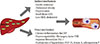
Non-alcoholic fatty liver disease (NAFLD) is a spectrum of progressive liver diseases, including simple steatosis, non-alcoholic steatohepatitis, fibrosis, and eventually cirrhosis, in the absence of excessive alcohol consumption [1] and has been considered a hepatic manifestation of metabolic syndrome. NAFLD is a marker of pathologic ectopic fat accumulation combined with a low-grade chronic inflammatory state [2]. The liver is a pivotal visceral organ with respect to fat accumulation. Accumulation of lipid droplets decreases the efficacy of insulin signaling. Hepatic lipids also induce endoplasmic reticulum stress, leading to activation of c-Jun N-terminal kinases and nuclear factor κB (NF-κB), two major regulators of inflammatory pathways that can inhibit phosphorylation of insulin receptor substraste-1 [3]. Recent evidence suggests that NAFLD is an emerging risk factor for cardiovascular disease (CVD) independent of traditional CVD risk factors, implying that NAFLD might be directly involved in CVD pathogenesis. Here, we will summarize the relationship between NAFLD and CVD, focusing on the causative roles of liver-derived hepatokines in the progression of CVD (Fig. 1).
EPIDEMIOLOGIC STUDIES LINKING NAFLD TO CVD
A recent comprehensive systematic review of 27 studies showed that NAFLD is associated with subclinical atherosclerosis independent of traditional risk factors and metabolic syndrome and across a wide range of ethnicities [4]. In that review, 14 among a total of 16 studies that examined the association of NAFLD with carotid intima media thickness (CIMT) demonstrated a significant increase in the mean CIMT in individuals with NAFLD compared to healthy people, even after adjusting for other confounding variables [4]. Using multislice computed tomography, Akabame et al. [5] showed that liver fat was significantly related to the presence of vulnerable plaques with a lipid core. Furthermore, Villanova et al. [6] reported that brachial artery flow-mediated dilatation (FMD), representing endothelial dysfunction, was reduced by 48% in subjects with NAFLD and that NAFLD predicted a reduced FMD (odds ratio [OR], 6.7; 95% confidence interval [CI], 1.26 to 36.1) after adjusting for age, sex, body mass index, and insulin resistance.
Several large cross-sectional and prospective studies showed that NAFLD was associated with an increased prevalence of CVD and the incidence of cardiovascular (CV) events irrespective of other CVD risk factors. In 2,839 patients with type 2 diabetes mellitus (DM), those with NAFLD had a higher prevalence of coronary, cerebrovascular, and peripheral vascular disease than their counterparts without NAFLD, independent of conventional CVD risk factors, medication history, and DM-related variables [7]. In addition, during a median 7.3 years of follow-up, an elevated baseline level of serum γ-glutamyl transferase, a surrogate marker for hepatic steatosis, was significantly associated with increased risk of all-cause and CVD mortality in men, even after adjusting for other metabolic risk factors [8]. Therefore, these studies suggested that assessment of NAFLD might be a helpful approach to more accurate and early CVD risk stratification.
MECHANISMS CONNECTING NAFLD WITH CVD
The underlying mechanism by which NAFLD increases the risk of CVD has not been clarified; moreover, whether NAFLD is an independent risk factor for CVD or simply a risk marker that coexists in people at increased risk of CVD is still controversial. The potential mechanism by which NAFLD increases CVD risk is based on the development of inflamed visceral adipose tissue [9], which is the main source of an elevated flux of free fatty acids (FFAs) into the portal vein for direct transport to the liver and subsequent hepatic fat accumulation [10]. Therefore, NAFLD can be considered a sensitive marker of pathological dysfunction of visceral adipose tissue that is more relevant to CV outcome than simply adipose tissue mass.
On the other hand, hepatic steatosis itself leads to intrahepatic inflammation through activation of NF-κB pathways that exacerbate insulin resistance both locally in the liver and systemically. The liver of subjects with NAFLD might release a variety of proatherogenic, proinflammatory, and diabetogenic mediators such as high-sensitivity C-reactive protein (hsCRP), fibrinogen, and plasminogen activator inhibitor-1 (PAI-1), which have important roles in the development of CVD [1]. hsCRP, which is primarily produced by the liver and is a marker of inflammation, was an independent predictor of CV events in several large studies [11]. Similarly, fibrinogen and PAI-1 also originate from hepatic tissue and are activators of the coagulation system thus enhancing atherothrombosis [2], suggesting that an increase in liver-secreted factors in NAFLD plays an important role in the pathogenesis of systemic inflammation and atherosclerosis. Therefore, the liver functions as an inducer of systemic inflammation as well as a target organ of various inflammatory reactions that occur within dysfunctional adipose tissue.
Recent evidence suggests that a group of predominantly liver-derived proteins called hepatokines directly affect glucose and lipid metabolism, similar to previously described adipokines and myokines [12]. At the present time, fetuin-A, fibroblast growth factor 21 (FGF-21), and selenoprotein P (SeP) are considered representative hepatokines; however, there have been very limited studies exploring the direct function of these hepatokines on the development of CVD.
FIBROBLAST GROWTH FACTOR 21
FGF-21 is a 181-amino acid peptide hormone that is primarily secreted by the liver and acts as a potent metabolic regulator [13]. Expression of the human FGF-21 gene is mediated by peroxisome proliferator-activated receptor-α (PPAR-α) during starvation [14] and regulated by PPAR-γ after feeding [15]. Circulating FFAs, a characteristic feature of fasting, and different kinds of stress such as hepatic injury, chemical insult, and disease, stimulate secretion of FGF-21 into the circulation [16]. Administration of FGF-21 to animal models and humans has been shown to decrease body weight and levels of blood triglycerides and low density lipoprotein (LDL) cholesterol, and to improve insulin sensitivity. Mashili et al. [17] reported that FGF-21 treatment induced basal and insulin-stimulated glucose uptake in human skeletal muscle cells through upregulation of glucose transporter-1. Very recently, a randomized phase 1 clinical trial showed that treatment with LY2405319, an analog of FGF-21, produced significant improvements in dyslipidemia of obese human subjects with type 2 DM [18]. FGF-21 stimulates lipolysis in the diabetic db/db mouse and obese humans, and individuals with a cluster of metabolic disorders have increased serum FGF-21 levels [19] to compensate for the abnormal metabolic status.
Early studies on FGF-21 focused on its role as a metabolic hormone during fasting or starvation. More recent studies have indicated a possible role of FGF-21 in CVD. Planavila et al. [20] demonstrated that FGF-21 knockout mice exhibited increased cardiac mass and impaired cardiac function, which could be ameliorated by treatment with FGF-21, suggesting its protective role against hypertrophic insults. Furthermore, FGF-21 infusion into a rat heart significantly recovered cardiac function following myocardial infarction [21]. In cultured rodent cardiac microvascular endothelial cells, FGF-21 expression was upregulated when the cells were incubated with oxidized LDL, indicating that FGF-21 might be secreted by endothelial cells in response to stress and that elevated levels may be a signal of endothelial cell injury [22]. In accordance with these animal studies, elevated serum FGF-21 levels are associated with carotid atherosclerosis in humans, independent of established CVD risk factors [23]. Multivariate logistic regression analysis also identified serum FGF-21 level as one of the independent factors of coronary artery disease occurrence (OR, 2.98; 95% CI, 1.014 to 8.786; P<0.05) [24]. Furthermore, we previously reported that brachial-ankle pulse wave velocity reflecting arterial stiffness had a significant positive correlation with circulating FGF-21 levels [25]. Therefore, FGF-21 may be an attractive target for the diagnosis and treatment of obesity and related diseases, including CVD.
FETUIN-A
Fetuin-A is a 64-kDa phosphorylated glycoprotein that is primarily synthesized by hepatocytes [26]. Fetuin-A is a natural inhibitor of the insulin receptor tyrosine kinase, leading to insulin resistance in rodents [27]. Apart from its direct effects on the insulin receptor, fetuin-A promotes insulin resistance by propagating a proinflammatory state. Fetuin-A treatment aggravates proinflammatory cytokine expression while reducing adiponectin expression in both adipocytes and monocytes [28]. Furthermore, incubation of HepG2 cells or rat hepatocytes with palmitate stimulates binding of NF-κB to the fetuin-A promoter, thereby augmenting fetuin-A synthesis and secretion [29]. We previously reported that palmitate-induced fetuin-A stimulated triacylglycerol accumulation in hepatocytes and that adiponectin inhibited palmitate-induced hepatic fetuin-A expression through the adenosine monophosphate-activated protein kinase (AMPK) pathway [30]. These results suggest that fetuin-A might directly cause insulin resistance and modulate inflammatory reactions, leading to various metabolic disturbances. Consistent with these findings, many epidemiologic studies have observed elevated levels of circulating fetuin-A in obesity and related metabolic diseases including type 2 DM, metabolic syndrome, and NAFLD [31,32,33]. Our previous study showed a significant decrease in circulating fetuin-A levels after 12 weeks of caloric restriction that was accompanied by improvements in visceral fat area, blood pressure, lipid profiles, and glucose levels [34]. In the European Prospective Investigation into Cancer and Nutrition-Potsdam study, plasma fetuin-A levels were positively associated with the incidence of diabetes after adjustment for sex, body mass index, waist circumference, and lifestyle risk factors during 7 years of follow-up [35].
However, the relationship between circulating fetuin-A and CVD risk appears to be more complicated. Fetuin-A can bind with Ca2+, inhibiting ectopic calcification [36]. In studies examining patients with chronic kidney diseases, fetuin-A level is inversely associated with calcification scores, CV events, and CV mortality [37]. Nondiabetics with a higher fetuin-A level have decreased risks of incident CVD and CVD-related mortality, whereas type 2 diabetics with higher fetuin-A levels have increased risks of incident CVD and CVD-related mortality [38,39]. Possible mechanisms by which fetuin-A can promote atherosclerosis in CVD patients are through induction of insulin resistance and increased expression of cytokines in monocytes that participate in the inflammation. Siegel-Axel et al. [40] demonstrated that fetuin-A inhibited the proliferation of perivascular fat cells and increased the expression of proinflammatory cytokines including interlukin-8, interlukin-6, monocyte chemotactic protein-1, and PAI-1 in these cells. Further studies should be performed to determine the exact function of fetuin-A in CVD according to the diverse underlying patient conditions.
SELENOPROTEIN P
SeP, a 42-kDa glycoprotein, is produced in the liver and secreted into plasma [41]. SeP was recently identified as a hepatokine associated with insulin resistance in humans through serial analysis of gene expression [42]. Administration of SeP to mice decreased insulin signaling and glucose metabolism in both liver and skeletal muscle, whereas SeP-deficient mice showed enhanced insulin signaling and improved glucose tolerance [42]. In our previous studies, patients with type 2 DM and those with NAFLD had higher serum SeP levels than healthy controls [43,44]. Furthermore, we found that salsalate and adiponectin ameliorated palmitate-induced insulin resistance in hepatocytes by inhibition of SeP through the AMPK-Forkhead box protein O1α (FOXO1α) pathway [45], suggesting that SeP acting via the AMPK-FOXO1α-dependent pathway might be a novel mechanism mediating the antidiabetic effects of salsalate and adiponectin.
However, there have been very few studies on the relationship of SeP with CVD. We first reported that circulating SeP level has an independent association with carotid intima-media thickness even after adjustment for other confounding factors [44]. Very recently, Ishikura et al. [46] showed that physiological concentrations of SeP inhibited vascular endothelial growth factor-stimulated cell proliferation, tubule formation, and migration in human umbilical vein endothelial cells, leading to impaired angiogenesis and delay of wound closure in mice overexpressing SeP. Further studies are needed to explore the direct relationship between SeP with CVD and the underlying mechanism.
CONCLUSIONS
Hepatokines that are mainly secreted from the liver are known to directly affect glucose and lipid metabolism. There is accumulating evidence that various hepatokines can modulate inflammatory processes that in turn mediate atherosclerotic process. Therefore, identification of novel hepatokines might allow the development of new strategies to diagnose and treat NAFLD-related metabolic disturbances, including CVD.
 XML Download