

 PDF
PDF ePub
ePub Citation
 Print
Print


INTRODUCTION
Diabetes is a complex condition characterized by chronic hyperglycemia and is associated with atherothrombotic complications. Atherothrombosis is the main cause of morbidity and mortality in patients with diabetes [1]. Diabetes increases the risk of coronary heart disease, stroke, and peripheral arterial disease by 2- to 4-fold [2]. The increased risk is independent of and additive to other cardiovascular disease (CVD) risk factors, such as hypertension, obesity, smoking, and dyslipidemia, relative to nondiabetic patients with these comorbidities [3]. In addition, patients with diabetes but without previous myocardial infarction (MI) have the same level of risk for subsequent acute coronary events as nondiabetic patients with previous MI. Diabetes has been classified as a coronary risk equivalent [4].
Diabetes has been considered to be a prothrombotic status. Several factors contribute to the prothrombotic condition as increasing coagulation, impaired fibrinolysis, endothelial dysfunction, and platelet hyperreactivity. Platelet plays a crucial role in the pathogenesis of atherothrombosis in diabetes [5]. A variety of platelet function abnormalities has been reported in vitro and in vivo [6,7]. Furthermore, patients with diabetes are at increased risk of not responding to standard antiplatelet treatments for acute coronary syndromes, leading to higher mortality and recurrent CVD [8].
This review focuses on an overview on the current status of knowledge of platelet function abnormalities in patients with diabetes, trying to address available clinical marker of platelet hyperreactivity in diabetes.
PLATELET ACTIVATION AND ATHEROTHROMBOSIS
Platelets are anuclear cell fragments derived from megakaryocytes that circulate in the bloodstream of humans for 7 to 10 days. Their primary function is to stop hemorrhage resulting from tissue trauma and vascular injury. Platelets sensitively respond to environmental change. Rapid platelet adhesion and aggregation at sites of tissue trauma and vascular injury are key events in maintaining normal hemostasis [9]. However, the same processes may also lead to thrombotic disorders-the formation of platelet plugs at sites of atherosclerotic lesion rupture is the most common mechanism leading to myocardial or cerebral infarction. Platelet activation plays important roles in both platelet adhesion and platelet aggregation. Suboptimal platelet activation or a hyperreactive state induced by different platelet signaling pathways may facilitate platelet adhesion and aggregation and accelerate thrombosis [10].
Platelet activation is initiated by the binding of thrombogenic substances, such as collagen, thrombin, or components of atheromatous plaque, to receptors located on the platelet surface [9]. Activated platelets can influence the progression of plaque formation by releasing adhesive ligands, such as P-selectin, that is expressed on the platelet membrane and mediates platelet-endothelium interactions. Signaling of P-selectin stimulates monocytes and macrophages to produce chemoattractants or growth factors [10].
Platelets are highly granular. Alpha granules contain a variety of adhesion molecules, chemokines, coagulation and fibrinolysis proteins, growth factors, and immunologic molecules. Dense granules contain ionic calcium, phosphate, magnesium, and serotonin, as well as adenosine triphosphate, guanosine triphosphate, adenosine diphosphate (ADP), and guanosine diphosphate nucleotides. As a result of activation, platelets release the content of their granules, which leads to autoactivation and amplification of aggregation and adhesion and accelerate the processes of vascular occlusion [11]. Adenosine monophosphate-activated protein kinase, protein kinase G, and protein kinase C are also involved in platelet degranulation by phosphorylating proteins involved in platelet secretion; such as Sec 1 and syntaxin 4 [12]. Activated platelet also releases inflammatory and mitogenic mediators into the local microenvironment, thereby altering the chemotactic, adhesive, and proteolytic properties of endothelial cells. Interaction of activated platelets with other cells, such as endothelial cells and leukocytes, may amplify the process of atherothrombosis [10].
Activated platelets release two types of membrane vesicles: platelet-derived microparticle (PMP) budded from the plasma membrane and exosomes, which are smaller than PMP and are released from α granules during secretion. Activated PMP also releases chemokines that can trigger the recruitment of monocytes or promote their differentiation into macrophages [13].
PLATELET FUNCTION ABNORMALITIES IN DIABETES
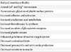
Platelets of diabetic patients are characterized by dysregulation of several signaling pathways and have been suggested to be hyperreactive, showing increased adhesion, activation, and aggregation [14]. Platelets from patients with type 1 and type 2 diabetes exhibit enhanced platelet activity early in the disease course that may precede the development of CVD [15]. Several mechanisms may account for the increased platelet activity in diabetes (Table 1). The glycation of platelet surface proteins reduces membrane fluidity and increases platelet adhesion, causing incorporation of glycated proteins into the thrombi. An increase in calcium mobilization from intracellular storage pools, resulting in increased intracellular calcium levels, has been correlated with reduction in membrane fluidity [16]. Platelet dysfunction in diabetes may be found even before development of visible damage to the vessel wall. Platelets in diabetes respond more frequently even to subthreshold stimuli, and thus contribute to accelerated thrombosis and release of fresh hyperreactive platelets [17].
Insulin has a direct inhibitory effect on platelet aggregation. Insulin binds to platelet membrane receptor and reduces platelet response to thrombin, ADP, arachidonic acid, collagen, and platelet activating factor. Diabetic platelets are less sensitive to the inhibitory action of insulin [18]. There is a decrease in platelet insulin receptor number and affinity in type 2 diabetes, which suggest that reduced insulin sensitivity may account for platelet hyperreactivity in this condition [19].
CLINICAL MARKER OF PLATELET HYPERREACTIVITY IN DIABETES
Mean platelet volume
Larger platelets are more active hemostatically and enzymatically, and they contain more prothrombotic molecules, such as platelet factor 4, serotonin, and platelet-derived growth factor, and possess greater aggregability in response to ADP [20]. Mean platelet volume (MPV), which is used to measure platelet size, can reflect platelet activity [21]. Increased MPV may lead to a prothrombotic condition with increased thromboxane A2 (TXA2) and B2 and adhesion molecule expression, such as P-selectin and glycoprotein IIb/IIIa, and β-thromboglobulin release [20].
Previous studies have shown that MPV is increased in diabetes and prediabetes [22-24]. Additionally, increased MPV has been associated with poor glycemic control in diabetes, the duration of diabetes, microalbuminuria, coronary heart disease, and an increase in the number of diabetic complications [25-27]. Increased MPV can be restored to normal level through improved glycemic control [20]. However, Shah et al. [28] reported a significant correlation between MPV and the degree of glycemic control only in diabetic patients. This result suggests that the positive relationship between an increased glucose level and increased MPV is a unique phenomenon of diabetes itself. Recent data from our group have supported this possibility. We found a contrasting relationship between MPV and fasting plasma glucose (FPG) in the presence and absence of diabetes in a Korean general population. After adjusting for confounding variables, MPV was only positively correlated with FPG in newly diagnosed diabetic women (β±SE, 0.097±0.037; P=0.016). Interestingly, MPV showed a significant inverse relationship with FPG in intermediate hyperglycemia (men: β±SE, -0.072±0.027, P=0.007; women: β±SE, -0.111±0.035, P=0.002) and normal glucose tolerance status (men: β±SE, -0.112±0.033, P<0.0001; women: β±SE, -0.102±0.034, P=0.003) [29]. The underlying mechanism behind this finding is unclear. Possible explanations include osmotic swelling due to increased blood glucose or other metabolites [30] and lack of a physiological inverse relationship between MPV and platelet count in diabetes, although increased platelet counts by increased glucose levels may lead to a subsequent decrease in MPV [20].
Thromboxane A2
Increased arachidonic acid metabolism in platelets from diabetic patients leads to enhanced TXA2 production and may contribute to increased platelet sensitivity [10]. Abnormalities in TXA2 production were among the earliest characterized abnormalities in platelet of diabetes [15]. The production of thromboxane can be measured by the urinary excretion of 11-dehydro-TXB2, its major enzymatic metabolite. Urinary 11-dehydro-TXB2 excretion was found to be significantly increased in type 2 diabetes [33]. Tight metabolic control has been shown to result in a significant reduction in 11-dehydro-TXB2 excretion [34].
Adhesion molecules and receptors
Hyperglycemia may induce nonenzymatic glycation of platelet membrane proteins with changes in protein structure and conformation, as well as alterations of membrane lipid dynamics [14]. Platelets from diabetes were found to have increased expression of adhesion molecules (CD31, CD36, CD49b, CD62P, CD63) and platelet surface receptors (GP Ib and GP IIb/IIIa), which mediate binding to von Willebrand factor in diabetes compared to controls [35]. After improving glycemic control for 3 months, a significant decrease in expression of CD31, CD49b, CD629, and CD63 was noted. Furthermore, a significant correlation between CD62P, CD63, and hemoglobin A1c (HbA1c) levels was also observed [36]. These findings suggest that improvement of glycemic control may have benefit on platelet adhesion and subsequent activation. The increased expression of GP IIb/IIIa is also consistent with enhanced fibrinogen binding and aggregability seen in platelets from diabetes [37].
Activated platelets can also release P-selectin, which is an adhesive ligand-mediating platelet-endothelium interactions. Since P-selectin on activated platelets has been shown to bind to CD15 or P-selectin glycoprotein ligand-1 expressed on monocytes and neutrophiles to form heterophilic aggregates, the increased P-selectin expression on platelets accounts for the increased formation of platelet-leukocyte aggregates in diabetes [10,15]. Levels of HbA1c and fasting glucose have been correlated to P-selectin expression in type 2 diabetes undergoing coronary angioplasty, which suggests that improved glycemic control may reduce platelet hyperreactivity [38].
CD40 ligand
Activated platelets release inflammatory and mitogenic mediators, altering the properties of endothelial cells [10]. CD40 ligand is stored in platelets and rapidly appears on the surface after platelet activation. Once released from platelets, CD40 induces inflammatory responses in the endothelium [39]. Increased plasma levels of CD40 ligand have been found in type 1 and type 2 diabetic patients [40]. Hyperglycemia and hyperinsulinemia induces increase of platelet derived CD40 ligand and monocyte-derived tissue factor [41]. In type 2 diabetes, circulating levels of CD40 ligand is correlated with progression of carotid intima-media thickness [42]. The significant correlation between plasma CD40 ligand level and urinary excretion of the 11-dehydro-TXB2 supports the likelihood of release of CD40 ligand during TXA2-dependent platelet activation in type 2 diabetes [43].
Platelet-derived microparticle
PMPs are veritable vectors for the intercellular exchange of biological signals and information; additionally, they may serve as shuttle modules and signaling tranducers not only in a certain local environment, but also at a remarkable distance from their site of origin since they readily circulate within vasculature [13]. PMP can bind to the exposed subendothelial matrix, thereby providing a substrate for further platelet adhesion via GP IIb/IIIa fibrinogen bridging. Circulating PMP also represent a phospholipase A2 substrate and lead to the production of lysophosphatidic acid, a strong platelet agonist [44]. Platelets and PMPs share glycoprotein receptors such as GPIb, PECAM-1, and integrin αIIbβ3, and subpopulations may express P-selectin from platelet granules, suggesting that PMP can participate in cellular interactions, adhesion, and aggregation [45]. Increased levels of PMPs have been reported in diabetic patients with macrovascular complications and have been suggested to play a role in these complications [46].
Reactive oxygen species
Diabetes is associated with increased oxidative stress, in particular with overproduction of reactive oxygen species (ROS), as well as reduced platelet antioxidant levels [15]. The major sources of ROS in platelets include nicotinamide adenine dinucleotide/nicotinamide adenine dinucleotide phosphate oxidase, superoxide-dismutase, and activated cyclooxygenase of phophoinositide metabolism. Activated platelets can generate ROS. The production of ROS by platelets enhances the recruitment of platelets to growing thrombus [47,48]. The platelet from type 2 diabetic patients showed enhanced endogenous ROS generation, increased intracellular Ca2+ and tyrosin phophorylation in response to thrombin in comparison with platelet from nondiabetes [15]. Increased ROS formation leads to enhanced lipid peroxidation and free radical-catalyzed conversion of arachidonic acid into bioactive F2-isoprostanes, such as 8-iso-prostaglandin F2α (PGF2α). The 8-iso-PGF2α is able to affect platelet functions as in vivo marker of oxidative stress [49]. Diabetic patients have higher levels of 8-iso-PGF2α, particularly in association with acute hyperglycemia episodes [50]. Vitamin E replacement in type 2 diabetes showed a significant reduction in urinary excretion of 8-iso-PGF2α [51].
CONCLUSIONS
Platelets are dynamic cells with activities that extend beyond primary hemostasis, including a pivotal role in initiating and progressing of atherothrombosis. Atherothrombotic complications are the important causes of morbidity and mortality in diabetic patients. Among the factors that contribute to the prothrombotic status in diabetes, abnormalities of platelet function play a crucial role. These platelet abnormalities ultimately result in a platelet hyperreactivity. Unfortunately, clinical markers of platelet hyperreactivity easily usable in clinical practice are lacking. MPV is universally available with routine blood counts by automated hemograms and therefore is an attractive index to evaluate. We suggest that MPV can be considered as an indicator of platelet function in clinical practice. Further prospective studies are needed.
Although understanding the platelet function abnormalities in diabetes still remains a challenge, more attention should be focused on platelet function for effective management and the prediction of atherothromboic events in diabetic patients.
 XML Download